Introduction
Pregnant women constitute 1.5 % of the patients who admitted to the hospital because of trauma [7]. It is known that prenatal blunt trauma may be a non-obstetric cause of maternal death, as well as may create prenatal intracranial hemorrhage such as subdural, epidural or intraparenchymal hemorrhage. In addition, many other etiological factors can cause this type of bleeding [3]. These patients are difficult to treat since they require many clinics to work together. We reported here a newborn suffered prenatal subdural hematoma and discussed possible reasons of hemorrhage with follow-up and treatment algorithm.
Case Presentation
A 25-years-old pregnant woman was treated with low-molecular weight heparin (LMWH) for habitual abortion beginning in the first week of gestation. She fell face down on the 35th week of pregnancy but she did not apply a hospital because she did not feel any serious discomfort. Vaginal bleeding and contractions occurred 10 days after trauma and she was immediately transported to an obstetric clinic.
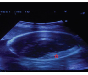
On the ultrasound, it was determined that there was a left frontoparietal lesion occupying space at fetus (fig. 1), due to vaginal bleeding and contraction and fetal maturation was appropriate, it was decided to perform cesarean section. At birth, the newborn’s body weight was 2620 g, and he was 52 cm long. The Apgar scores were 7 and 9 at 1 and 5 minutes. After birth, the mild right hemiparesis was present and during the early postnatal hours moderately reactive and no oral intake. Shortly after birth, coagulation parameters of newborn was checked and found to be within normal limits. (PT; 24.9 sec, aPTT; 29 sec, INR; 1.91, PLT; 215.000, Factor XII; 185 %, Factor IX; 41 %, Factor VII; 22 %, Factor VIII; 400 %, Fibrinogen; 508 mg/dl, protein C 21 %, protein S 57 %, Homocysteine; 4.49 umol/l).
/85.jpg)
A postnatal brain computed tomography and magnetic resonance imaging studies confirmed the large left hemispheric subdural hematoma (Lesion appears isointense on T1-weighted image and mix-intensity on T2-weighted image. Septa were identified within the hematoma cavity by MRI, maximum thickness were 18.57 mm) that caused mass effect and 1 cm midline shift (fig. 2).
/85_2.jpg)
After the newborn’s vital signs was stabilized, the newborn was undergoing neurosurgical procedure. We planned and applied frontoparietal craniotomy, as the hematoma was thought to be solid component according to the tomography. The hemorrhage collection which consisted of a scarce fluid component and a large blood clot attached to thick parietal membranes, was completely removed (fig. 3). The newborn was transferred neonatal intensive care unit for postoperative intensive treatment. The newborn was discharged to the pediatric department at 7th days after surgery in good clinical condition. The postoperative tomography demonstrated that evacuation of subdural hematoma and no residual collection.
Discussion
One of the reasons that endanger the life of mother and fetus during pregnancy in trauma. It is rare and important cause of morbidity and mortality. Trauma has many varieties in pregnancy and the most common ones are motor vehicle accidents and falls [3].
Therefore, brain pathologies such as intracranial hemorrhages, cranial fractures and hypoxic ischemic encephalopathy can be seen in fetuses of third trimester pregnant women exposed to blunt abdominal trauma. There is no definite incidence of intrauterine intracranial hemorrhages. In addition, there are no specific clinical findings. It is asymptomatic or there are nonspecific clinical signs such as decreased fetal movements. The diagnosis is made after ultrasonographic imaging performed in pregnant women who come with a symptomatic or asymptomatic clinic. There are several etiological causes of intrauterine intracranial hemorrhage such as trauma, perinatal asphyxia, drug use affecting platelet activity, infection, and congenital factor deficiency. However, there is a closer relationship between intrauterine subdural hematomas and maternal trauma than other etiological causes [5, 6].
We found trauma as an etiological cause. Although it is stated in a publication that there is an intrauterine subdural hematoma after the use of LMWH, it is known that LMWH does not pass through the placenta [1, 4].
Subdural hematoma is rare in newborns. It is rarely observed in antenatal period. This is the result of the protective efficacy of the maternal abdominal and pelvic structures and amniotic fluid. In a study conducted by Joseph et al in 2017 with PubMed publications, a total of 14 cases have been reported to date. Only 4 of these cases were isolated subdural hematoma [3, 5].
Ultrasound imaging (USG) and Magnetic resonance imaging (MRI) are effective and safe methods of diagnosis in antenatal brain hemorrhages. USG is essential for screening and diagnosis in antenatal intracranial hemorrhages. However, Fetal USG may give false information in some cases such as fetal head position, hemorrhage location and small hematoma size. Therefore, in suspicious cases, CT and MRI can be used to better view the brain tissue. MRI use is limited due to several reasons such as claustrophobia and long stay. CT is an important imaging method that provides information for both the maternal and the fetus in clinical indication. In addition, bone tissues are better evaluated [8].
The management of the patient diagnosed with intrauterine intracranial hemorrhage requires a multidisciplinary perspective. Obstetric intervention, intracranial hemorrhage and general condition of the fetus should be evaluated together. Urgent intervention or follow-up decision should be made by gynecology, neurosurgery, and pediatrics. In cases where the bleeding size is small, lung maturation does not develop, and the fetus is clinically stable, obstetric intervention may be delayed. USG monitoring should be performed frequently in such patients. In case of clinical suspicion, additional imaging tests such as MRI and CT scan should be used [2].
If the size of intracranial hemorrhage is large, emergency obstetric intervention should be considered. Although it is stated that obstetric intervention is not an obstacle for vaginal delivery, we think that cesarean should be performed to the mother, which is the least traumatic method for the fetus [3].
CT and MRI tests should be performed early in the newborn period. These imaging methods are important for the visualization of bleeding area, bone tissue, brain parenchyma and ischemic areas. In addition, these imaging methods are important in terms of morbidity estimation and family information. Newborn should be treated in neonatal intensive care in terms of post-surgical care and hematological tests [8].
Conclusions
Intrauterine subdural hematoma is very mortal and rare. The etiological cause should be investigated well. Neurosurgery, radiology, and neonatal intensive care clinics are required for obstetric intervention type and timing and diagnosis and treatment of newborn. Pregnant should be treated from a multidisciplinary perspective.
Received 07.08.2021
Revised 17.08.2021
Accepted 20.08.2021
Список литературы
1. Bauder F., Beinder E., Arlettaz R., Albisetti M., Boltshauser E., Gessler P. Intrauterine subdural hemorrhage in a preterm neonate possibly associated with maternal low-molecular weight heparin treatment. J. Perinatol. 2009. 29. 521-523.
2. de Spirlet M., Goffinet F., Philippe H.J., Bailly M., Couderc S., Nisand I. Prenatal diagnosis of a subdural hematoma associated with reverse flow in the middle cerebral artery: case report and literature review. Ultrasound Obstet. Gynecol. 2000. 16. 72-76.
3. Green-Thompson R., Moodley J. In-utero intracranial haemorrhage probably secondary to domestic violence: case report and literature review. J. Obstet. Gynaecol. 2005. 25. 816-818.
4. Greer I.A., Brenner B., Gris J.C. Antithrombotic treatment for pregnancy complications: which path for the journey to precision medicine? Br. J. Haematol. 2014. 165. 585-599.
5. Joseph J.R., Smith B.W., Garton H.J. Blunt prenatal trauma resulting in fetal epidural or subdural hematoma: case report and systematic review of the literature. J. Neurosurg. Pediatr. 2017. 19. 32-37.
6. Piastra M., Pietrini D., Massimi L., Caldarelli M., De Luca D., Del Lungo L.M., De Carolis M.P., Di Rocco C., Conti G., Zecca E. Severe subdural hemorrhage due to minimal prenatal trauma. J. Neurosurg. Pediatr. 2009. 4. 543-546.
7. Sakamoto J., Michels C., Eisfelder B., Joshi N. Trauma in Pregnancy. Emerg. Med. Clin. North Am. 2019. 37. 317-338.
8. Zeina A.R., Kessel B., Mahamid A., Gazmawi J., Shrim A., Nachtigal A., Alfici R. Computed tomographic diagnosis of traumatic fetal subdural hematoma. Emerg. Radiol. 2013. 20. 169-172.

/85_2.jpg)
/86.jpg)