Кістозні захворювання нирок у дорослих — це гетерогенна група захворювань, що характеризуються наявністю множинних кіст у нирках [1]. Ці захворювання класифікують на спадкові, набуті або кістозні хвороби, що розвиваються, на підставі їх патогенезу. Спадкові захворювання включають автосомно-домінантний полікістоз нирок, медулярно-кістозну хворобу нирок, хворобу фон Гіппеля — Ліндау і туберозний склероз. Набуті стани включають кістозну хворобу нирок, що розвивається в пацієнтів з термінальною стадією ниркової недостатності. Кістозні захворювання нирок, що розвиваються, включають локалізовану кістозну хворобу нирок, мультикістозну диспластичну нирку й медулярну губчату нирку. Останніми роками виявлено багато молекулярних і клітинних механізмів, що беруть участь у патогенезі кістозних захворювань нирок [2–4]. Спадкові кістозні захворювання нирок характеризуються генетичними мутаціями, які призводять до дефектів у структурі й порушення функції первинних війок епітеліальних клітин ниркових канальців, аномальної проліферації епітелію канальців і підвищеної секреції рідини, що в кінцевому підсумку призводить до розвитку кіст нирки. Краще розуміння цих патофізіологічних механізмів на сьогодні є основою для розробки більш цільового лікування цих розладів. Візуалізація поперечного зрізу надає корисну інформацію щодо діагностики, спостереження, прогнозування й оцінки відповіді на лікування при кістозних захворюваннях нирок [1, 5, 6]. Диференціальну діагностику кістозних захворювань нирок подано в табл. 1.
/82.jpg)
Мультикістозна диспластична нирка (МКДН) — це дисплазія нирок, що характеризується наявністю множинних, не сполучених між собою кіст різного розміру, розділених диспластичною паренхімою, які поглинають кору нирки, що призводить до розвитку нефункціонуючої нирки з відсутністю нормальної архітектоніки чашко-мискової системи. Це рідкісна вада розвитку, яка за класифікацією належить до вад розвитку структури ниркової паренхіми, що пов’язано з атрезією системи зачатків сечоводу на метанефральній стадії розвитку матки з порушенням нормального нефрогенезу [7]. Захворюваність на МКДН становить приблизно 1 : 4300 новонароджених, при цьому чоловіки хворіють частіше, ніж жінки. МКДН може бути сімейним захворюванням, але найчастіше виникає спорадично. Більшість випадків МКДН виявляються під час ультразвукового дослідження плода, і про них повідомляють вже на 15-му тижні вагітності. Дослідження показали, що МКДН виникає у 53 % випадків у лівій нирці й у 47 % випадків — у правій нирці [7–9].
МКДН може бути односторонньою, двосторонньою або сегментарною і може додатково класифікуватися як проста чи складна. Проста МКДН визначається як одностороння дисплазія з нормальною контралатеральною ниркою, але з компенсаторною гіпертрофією контралатеральної нирки і без супутніх сечостатевих аномалій. Складна МКДН визначається як двостороння дисплазія контралатеральної нирки або сечостатеві аномалії. МКДН можуть бути пов’язані з низкою інших дефектів, що включають ротаційні або позиційні аномалії, гіпоплазію, ділянку дисплазії, міхурово-сечовідний рефлюкс, уретероцеле, обструкцію мисково-сечовідного сегмента. Міхурово-сечовідний рефлюкс є найбільш поширеною нирковою аномалією в пацієнтів із МКДН, що зустрічається приблизно у 25 % випадків. Двостороння МКДН несумісна з життям [10–12].
При односторонньому ураженні мультикістозна нирка може не мати клінічної симптоматики й бути випадковою знахідкою.
Клінічний випадок гідронефротичної форми мультикістозної дисплазії нирки, що ускладнилась септичним пієлонефритом, у дорослої людини
Пацієнтка М., 40 років, звернулась на консультацію зі скаргами на лихоманку до 40 °С, нудоту, блювання, наростаючу загальну слабкість, неприємні відчуття в правому боці. Вищезазначені скарги турбують упродовж тижня, самостійно приймала антибіотик ципрофлоксацин 1 г на добу, знеболюючі, антипіретики. Без належного ефекту.
З анамнезу життя відомо: 10 років тому пацієнтка була госпіталізована зі схожими скаргами, діагностовано кісти нирок, стеноз пієлоуретерального сегмента. Була проведена люмботомія, пластика пієлоуретерального сегмента. Виписана, зі слів пацієнтки, у стабільному стані. Після чого упродовж 10 років не обстежувалась, не лікувалась.
Об’єктивно: стан середньої тяжкості — тяжкий, продуктивному контакту пацієнтка доступна, проте на питання відповідає дещо із затримкою. Шкіра бліда. Пальпаторно болючість у правій здухвинній ділянці. Симптом подразнення поперекових м’язів праворуч позитивний. Оцінка показників життєдіяльності: артеріальний тиск 100/70 мм рт.ст., частота серцевих скорочень — 110 уд/хв, частота дихальних рухів — 25/хв.
При ультразвуковому дослідженні: права нирка значно збільшена в розмірах, має численні кістозні порожнини, максимальний розмір до 8,5 × 8,3 см. Уміст кістозних порожнин — ехо-позитивний. Кортико-медулярна диференціація не простежується. Нирковий синус і паренхіма не диференціюються (рис. 1, 2). Ліва нирка збільшена в розмірах, її об’єм становить 410 см3. Кортико-медулярна диференціація збережена. Товщина паренхіми 26 мм. Чашко-мисковий комплекс — без особливостей.
Дані лабораторних обстежень подано в табл. 2.
З огляду на те, що пацієнтка упродовж 5 діб приймала антибіотик ципрофлоксацин у дозі 1 г, бактеріологічне дослідження крові й сечі є недоцільним.
З урахуванням скарг, даних анамнезу й обстежень був встановлений попередній діагноз: гідронефротична форма мультикістозної дисплазії правої нирки. Ускладнений обструктивний пієлонефрит. Уросепсис.
Пацієнтці в терміновому порядку проведена черезшкірна перкутанна нефростомія справа під ультразвуковим і рентгеноскопічним контролем. Під час і після операції ускладнень не зареєстровано. За добу по дренажу евакуйовано до 1000 мл гнійного вмісту, який досліджено бактеріологічно. Виявлено комбінацію збудників, а саме P.aeruginosa 107 КУО/мл і Enterococcus faecalis 105 КУО/мл.
Згідно з керівництвами EAU з лікування урологічних інфекцій 2022 року, препаратами вибору для лікування ускладнених інфекцій сечових шляхів із системними симптомами є комбінація аміноглікозидів і В-лактамних антибіотиків (рис. 3) [13].
Призначена терапія: гентаміцин 80 мг внутрішньом’язово 2 рази на добу, цефтріаксон в/в краплинно 4 г на добу. Дезінтоксикаційна і симптоматична терапія.
Уже на другу добу після нефростомії і початку антибактеріальної терапії стан пацієнтки мав значну позитивну динаміку: регрес симптомів інтоксикації, максимальна температура тіла не перевищувала 37,8 °С, по стомі — гнійний вміст. На 10-ту добу лікування — стан пацієнтки задовільний, скарг не має. Температура тіла стабільно нормальна, повний регрес інтоксикаційного й больового синдромів. Динаміка лабораторних показників подана в табл. 3.
/85.jpg)
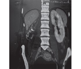
Пацієнтці проведена комп’ютерна томографія нирок з уведенням контрастної речовини. Стан після правобічної нефростомії. У правій нирці — множинні кістозні структури. Ниркова паренхіма витончена до 2 мм, архітектоніка порушена, відзначається слабке нерівномірне контрастування паренхіми нирки. Функція нирки не простежується. Сечовід не візуалізується. Надходження контрастної речовини до чашко-мискової системи й сечоводу немає. При проведенні ангіографії ниркових судин виявлено: права ниркова артерія відходить від черевного відділу аорти в типовому місці, діаметр 1,5 мм, аберантна нижньополярна ниркова артерія справа відходить нижче на 60 мм по передній частині аорти, діаметр 1,3 мм, і живить нижній полюс правої нирки. Ліва нирка та її ангіоархітектоніка — без особливостей (рис. 4–7).
Заключний клінічний діагноз: гідронефротична форма мультикістозної дисплазії правої нирки. Ускладнений обструктивний пієлонефрит. Уросепсис.
Вроджена аномалія розвитку судин правої нирки: аберантна нижньополярна ниркова артерія справа. Вазоренальний конфлікт. Стеноз мисково-сечовідного сегмента правої нирки.
Виписана із стаціонару в задовільному стані з рекомендаціями щодо подальшого хірургічного лікування — нефректомії правої нирки через 1 місяць.
У подальшому в плановому порядку після передопераційної підготовки виконана лапароскопічна нефректомія справа. Час операції становив 68 хв. Рівень інтраопераційної крововтрати — 20 мл. Інтра- і післяопераційних ускладнень не зареєстровано.
Гістологічне дослідження: макропрепарат — нирка розміром 12 × 8,5 × 3,5 см, на поверхні тканина неоднорідна, сіро-багряного кольору, з поверхні на більшому протязі визначаються множинні кісти діаметром від 0,2 до 1,5 см сірого кольору, на розрізі вміст серозний, з домішкою пластових мас, стінка гладка, сірого кольору. На розрізі паренхіма різко витончена до 0,2 см, практично не простежується, сіро-білого кольору, визначаються множинні, кістоподібні порожнини, у діаметрі від 1 до 8 см, внутрішня стінка кіст гладка, сірого кольору, заповнена серозним вмістом, з жовтуватим відтінком.
Мікропрепарат: у досліджуваних препаратах, забарвлених гематоксилін-еозином, тканина нирки представлена безліччю кіст різної величини, розташованих по всій тканині нирки, вистелених сплощеним епітелієм. Відзначаються дрібні ділянки з тканини нирки, що зберіглася, з невеликою кількістю клубочків і канальців. У стромі лімфогістіоцитарна інфільтрація. Миска з осередками розростання сполучної тканини, лімфогістіоцитарною інфільтрацією.
Висновок: мультикістозна дисплазія правої нирки.
Обговорення
МКДН зазвичай перебігає безсимптомно й тривалий час може залишатися недіагностованою, до дорослого віку. Більшість людей, які мають МКДН і одну нормально функціонуючу нирку, можуть вести нормальне здорове життя. Мультикістозна диспластична нирка може зберігатися без будь-яких змін, збільшення розмірів або навіть піддаватися спонтанній інволюції [14]. У нашому клінічному випадку в пацієнтки кісти нирок були діагностовані в дорослому віці, при першому епізоді інфекції сечових шляхів (ІСШ).
Деякі дослідження демонструють, що дорослих з неускладненою МКДН краще лікувати консервативно [15]. Існує багато суперечливих даних щодо можливості злоякісного переродження МКДН. Більшість повідомлень про такі випадки були пов’язані з пухлиною Вільмса, нирково-клітинним раком і уротеліальним раком, які розвинулися в пацієнтів із МКДН [12]. Хірургічне втручання при дисплазії нирки потрібне лише тоді, коли збільшена нирка не регресує, викликає обструктивну уропатію, розвивається злоякісне новоутворення або дисплазія нирки стає причиною артеріальної гіпертензії або ІСШ [16]. Оскільки частота ускладнень, таких як артеріальна гіпертензія, злоякісні новоутворення або ІСШ, у випадку МКДН невелика, потреба в хірургічному лікуванні дисплазії нирки виникає доволі рідко. Однак у деяких дослідженнях припускають, що нефункціонуюча МКДН може бути видалена, щоб уникнути ризику розвитку гіпертензії, злоякісного новоутворення та інших ускладнень, однак сама єдина нирка теж є фактором ризику через збільшення частоти контралатеральних аномалій [17]. У нашому клінічному випадку тільки консервативне ведення було неможливим унаслідок обструктивного септичного пієлонефриту нефункціонуючої диспластичної нирки, коли тільки за першу добу після нефростомії було виділено до 1000 мл гнійного вмісту. Крім того, вікарна гіпертрофія лівої нирки вірогідно вказує на давність процесу, тому епізод пієлонефриту МКДН є фактором ризику розвитку хронічного уросепсису й погіршення функції єдиної функціонуючої нирки до термінальних стадій і необхідності замісної ниркової терапії. Тому подальша нефректомія нефункціонуючої МКДН стала необхідним етапом лікування.
У пацієнтів з МКДН було описано безліч супутніх аномалій сечовивідних шляхів. Найчастішим і потенційно значущим урологічним дефектом є міхурово-сечовідний рефлюкс контралатеральної нирки. Також часто спостерігаються інші аномалії сечовивідних шляхів, такі як обструкція контралатерального мисково-сечовідного сегмента [18]. Наша пацієнтка 10 років тому була прооперована з приводу стенозу мисково-сечовідного сегмента диспластичної нирки, що розвинувся на тлі недіагностованого вазоренального конфлікту — аберантної нижньополярної ниркової артерії справа. Упродовж наступних 10 років вазоренальний конфлікт продовжував бути причиною стенозу мисково-сечовідного сегмента, що тільки погіршувало нормальний пасаж сечі через диспластичну нирку і в кінцевому результаті призвело до гідронефротичної трансформації МКДН з обструктивним септичним пієлонефритом і необхідності нефректомії.
Висновки
Хоча МКДН є рідким захворюванням, скринінгові ультразвукові дослідження можуть запобігти нирковим ускладненням. Крім того, необхідно проводити скринінг на інші супутні аномалії сечових шляхів як у нирці з мультикістозною дисплазією, так і контралатеральній нирці.
Пацієнти з наявністю множинних кіст (3 і більше без обтяженої спадковості та будь-яка кількість кіст у пацієнтів із сімейною історією) обов’язково мають проходити генетичне тестування на мутації генів PKD1, PKD2, PKHD1, MUC1 і MOD.
Пацієнти з МКДН можуть лікуватися консервативно в разі неускладненого перебігу, а питання щодо нефректомії нефункціонуючої МКДН має вирішуватися індивідуально, з урахуванням клінічних даних пацієнта й оцінки подальших прогнозів якості й тривалості життя.
Конфлікт інтересів. Автори заявляють про відсутність конфлікту інтересів і власної фінансової зацікавленості при підготовці статті.
Отримано/Received 28.05.2022
Рецензовано/Revised 09.06.2022
Прийнято до друку/Accepted 15.06.2022


/82.jpg)
/83.jpg)
/84.jpg)
/85.jpg)
/85_2.jpg)
/86.jpg)