
Архив офтальмологии Украины Том 11, №1, 2023
Вернуться к номеру
Запальна оптична нейропатія і атрофія зорового нерва (клінічні випадки)
Авторы: Мойсеєнко Н.М.
Івано-Франківський національний медичний університет, м. Івано-Франківськ, Україна
Рубрики: Офтальмология
Разделы: Справочник специалиста
Версия для печати
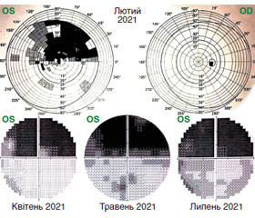
Актуальність. Класифікація, патогенез і клінічні прояви невриту зорового нерва неоднозначні. Привертає увагу складність статистичного аналізу подібних випадків, особливо на початковому етапі, коли прояви схожі, але трактуються по-різному, обираються різні лікувальні тактики. Мета: дослідити особливості атрофії зорового нерва, викликаної запаленням зорового нерва, за допомогою оптико-когерентної томографії (ОКТ). Матеріали та методи. Обстежено в амбулаторних умовах в Івано-Франківському національному медичному університеті 2 пацієнти (4 ока) із запаленням зорового нерва. Проведено візометрію, офтальмоскопію і аналіз даних комп’ютерної периметрії та ОКТ, виконаних у приватних установах у містах первинного обслуговування і в Івано-Франківську. Результати. Було обстежено два пацієнти, В., 31 рік, і К., 25 років, з прогресивним зниженням зору впродовж двох років і семи місяців. В обох випадках встановлено діагноз атрофії зорового нерва, асоційованої із вірусною інфекцією. У випадку пацієнта В. атрофія почалася зі збільшення товщини шару нервових волокон (ШНВ) на стороні ураження на 19 % порівняно із протилежною стороною, а в пацієнтки К. — відразу зі зменшення вдвічі. Надалі у першому випадку зменшилась товщина ШНВ і шару гангліонарних клітин (ШГК) вдвічі порівняно із попереднім періодом, а в другому випадку залишилась на попередньому рівні. Зменшення товщини ШНВ і ШГК може вважатися критерієм, що ускладнює прогноз при невриті, і може розглядатися як параметр трансформації запальної невропатії в атрофію зорового нерва. Висновки. Неврит є важливим фактором формування атрофії зорового нерва. Раннє застосування ОКТ-параметрів структурних пошкоджень головки зорового нерва дозволить диференціювати тяжкість ураження при запальній невропатії різного генезу.
Background. Classification, pathogenesis and clinical manifestations of optic neuritis are ambiguous. The complexity of the statistical analysis in such cases draws attention, especially at the initial stage, when the manifestations are similar, but are considered differently, various treatments are adopted. The purpose is to investigate the signs of optic nerve atrophy caused by inflammation of the optic nerve using optical coherence tomography (OCT). Materials and methods. Two patients (4 eyes) with inflammation of the optic nerve were examined in outpatient conditions at the Ivano-Frankivsk National Medical University. Visometry, ophthalmoscopy and analysis of computer perimetry and OCT data conducted in private institutions of the city were carried out. Results. Two patients were examined for over 2 years and 7 months: V. aged 31 years and K. aged 25 years with progressive vision loss. In both cases, the diagnosis was as follows: optic nerve atrophy associated with a viral infection. In patient V., the atrophy began with an increase in the thickness of the retinal nerve fiber layer (RNFL) by 19 % on the side of the lesion compared to the opposite side, and in patient K., it immediately decreased by half. Subsequently, in the first case, the thickness of the RNFL and retinal ganglion cell layer decreased twice compared to the previous period, and in the second case, it remained at the previous level. A decrease in the thickness of the RNFL and retinal ganglion cell layer can be considered a criterion that complicates the prognosis in neuritis and can be considered as a predictor for the transformation of inflammatory neuropathy into optic nerve atrophy. Conclusions. Neuritis is an important factor in the formation of optic nerve atrophy. Early use of OCT parameters of structural damage to the optic nerve head will allow differentiating the severity of damage in inflammatory neuropathy of various genesis.
неврит зорового нерва; запальна нейропатія; оптико-когерентна томографія; шар нервових волокон; атрофія зорового нерва
optic neuritis; inflammatory neuropathy; optical coherence tomography; nerve fiber layer; optic nerve atrophy
Для ознакомления с полным содержанием статьи необходимо оформить подписку на журнал.
- Costello F. Inflammatory Optic Neuropathies. Neuro-ophthalmology. 2014. 20 (4). 816-837.
- Yassa E.T., Bakbak B. Chronic Relapsing Inflammatory Optic Neuropathy. Med. Bull. Sisli. Etfal. Hosp. 2019. 53(4). 437-440.
- Wen Y.T., et al. Inhibition of Retinal Ganglion Cell Loss By a Novel ROCK Inhibitor (E212) in Ischemic Optic Nerve Injury Via Antioxidative and Anti-Inflammatory. Actions. Invest. Ophthalmol. Vis. Sci. 2021. 62 (6). 21.
- Moyseyenko N.M. The pathogenic aspects of traumatic optical neuropathy’s neuroprotec-tive therapy. Art of Medicine. 2019. 4 (12). 84-91.
- Sharma P., Sharma R. Toxic optic neuropathy. Indian. J. Ophthalmol. 2011. 59. 137-141.
- Chan J.W. Optic Nerve Disorders Diagnosis and Management. Springer is part of Springer Science: Business Media, 2014. 376.
- Grzybowski A., Barboni P. OCT and Imaging in Central Nervous System Diseases. The Eye as a Window to the Brain. Springer International Publishing AG Switzerland is part of Springer Science: Business Media, 2020. 343.
- Kidd D.P. Chapter 7 Inflammatory Optic Neuropathies Not Associated with Multiple Sclerosis. Blue Books of Neurology. 2008. 32. 153-190.
- Clare G., Colley S., Kennett R., Elston J.S. Reversible optic neuropathy associated with low-dose methotrexate therapy. J. Neuroophthalmol. 2005. 25 (2). 109-112.
- Sbeity Z.H., Baydoun L., Schmidt S., Loeffler K.U. Visual field changes in methotrexate therapy. Case report and review of the literature. J. Med. Liban. 2006. 54 (3). 164-167.
- Han S., et al. Altitudinal Visual Field Defect in NAION and OAG. Korean. J. Ophthalmol. 2015. 29 (6). 418-423.
- Masoud Aghsaei Fard, Hossein Ghahvehchian, Prem S. Subramanian. Optical coherence tomography in ischemic optic neuropathy. Ann. Eye Sci. 2020. 5. 6.
- Fraser J.A., Rueløkke L.L., Malmqvist L., Hamann S. Pre–valence of Optic Disc Drusen in Young Patients With Nonarteritic Anterior Ischemic Optic Neuropathy: A 10-Year Retrospective Study. J. Neuroophthalmol. 2021. 41(2). 200-205.
- Sanfilippo F., La Rosa V., Oliveri F., Astuto M. COVID-19, Hypercoagulability, and Cautiousness with Convalescent Plasma. American Journal of Respiratory and Critical Care Medicine. 2021. 203 (2). 257-258.
- Yang J.M., Park S.W., Ji Y.S., Kim J., Yoo C., Heo H. Postural effects on intraocular pressure and ocular perfusion pressure in patients with non-arteritic anterior ischemic optic neuropathy. BMC Ophthalmol. 2017. 17 (1). 47.