Інформація призначена тільки для фахівців сфери охорони здоров'я, осіб,
які мають вищу або середню спеціальну медичну освіту.

Підтвердіть, що Ви є фахівцем у сфері охорони здоров'я.

Международный неврологический журнал Том 20, №3, 2024

Вернуться к номеру

Нервово-м’язове з’єднання при бічному аміотрофічному склерозі. Що ще треба знати?

Авторы: Sh. Vashadze (1), M. Kekenadze (2, 3), N. Kvirkvelia (4), M. Beridze (2, 3)
(1) - Batumi Shota Rustaveli State University, Batumi, Georgia
(2) - National Hospital for Neurology and Neurosurgery, London, UK
(3) - The First University Clinic of Tbilisi State Medical University, Tbilisi, Georgia
(4) - Petre Sarajishvili Institute of Neurology, Tbilisi, Georgia

Рубрики: Неврология

Разделы: Клинические исследования

Версия для печати


Резюме

Актуальність. Бічний аміотрофічний склероз (БАС) викликає прогресуючу дегенерацію верхніх мотонейронів у корі головного мозку та нижніх мотонейронів у хребті. Крім того, незрозуміло, де починається дисфункція моторних нейронів і що викликає їхню дегенерацію. Це все ще є предметом дискусій. Матеріали та методи. Чи зустрічаються порушення нервово-м’язового зв’язку на ранніх стадіях БАС? Щоб відповісти на це питання, ми обстежили 5 пацієнтів із БАС (3 чоловіків і 2 жінки віком 50–61 рік) в Інституті неврології ім. Петра Сараджишвілі в 2018–2022 роках. Діагноз БАС ґрунтувався на клінічних ознаках, критеріях Gold Coast, результатах електроміографії (критерії Awaji), нейровізуалізації, аналізів крові та сечі. На ранній стадії захворювання спостерігалися лише асиметричний птоз і диплопія, які не покращувалися під впливом піридостигміну чи стероїдів. Результати. Вивчали анамнез пацієнтів, фізикальні дані, оцінювали психічні, когнітивні функції та неврологічний статус. Ми також опитували членів родини, оскільки пацієнту часто було важко точно описати симптоми. Антитіла до рецепторів ацетилхоліну були помірно позитивними лише в одного пацієнта. Тимому було виключено. При нейрофізіологічному дослідженні виявлено лише виражену недостатність нервово-м’язової передачі кругового м’яза ока, клінічних та електроміографічних ознак ураження мотонейронів не зафіксовано. Висновки. Приблизно через 2 роки у всіх п’яти пацієнтів розвинулися клінічні та електроміографічні ознаки БАС. У цьому дослідженні виявлено, що розлади нервово-м’язового з’єднання відіграють важливу роль у патогенезі БАС і можуть слугувати корисним раннім діагностичним маркером.

Background. Amyotrophic lateral sclerosis (ALS) causes progressive degeneration of upper motor neurons in the cortex, and lower motor neurons in the spine. In addition, it is unclear where motor neuron dysfunction begins and what causes motor neuron degeneration: whether it is the dying forward process or the dying back phenomenon where motor neuron degeneration begins distally at the nerve terminal or at the neuromuscular junction and progresses toward the cell body, is still a matter of debate. Materials and methods. Are there neuromuscular junction disorders in the early stages of ALS? To answer this, we described 5 patients with ALS presented at Petre Sarajishvili Institute of Neurology in 2018–2022, 3 males and 2 females aged 50–61 years. ALS diagnosis was based on clinical signs, the Gold Coast criteria, electromyography (Awaji), neuroimaging, blood and urine tests. At the early stage of the disease, only asymmetric ptosis and diplopia were noted, which did not improve on pyridostigmine or steroids. Results. We studied patients’ anamnesis, physical data, evaluated their mental, cognitive functions and neurological status. We have also interviewed family members, as it was often difficult for the patient to accurately describe the symptoms. Acetylcholine receptor antibodies were mildly positive only in one patient. Thymoma was excluded. The neurophysiological study showed only marked neuromuscular transmission failure in orbicularis oculi, there were no clinical and electromyographic signs of motor neuron damage. Conclusions. Approximately 2 years later, all five patients developed clinical and electromyographic signs of ALS. In the present study, neuromuscular junction disorders are found to play an important role in the pathogenesis of ALS and may serve as a useful early diagnostic marker.


Ключевые слова

бічний аміотрофічний склероз; розлади нервово-м’язового з’єднання; Грузія

amyotrophic lateral sclerosis; neuromuscular junction disorders; Georgia

Introduction

As of 2017, the Amyotrophic Lateral Sclerosis (ALS) Registry has found up to 31,843 cases of ALS in the United States with a mean of 24,821 and a lower estimate of 17,800. ALS is not a reportable disease in most states and CDC/ATSDR is not notified of these cases at this time. ALS is slightly more common in men than in women. ALS is age related; most people find out they have it when they are between 55 and 75 years of age and live from 2 to 5 years after symptoms develop. How long a person lives with ALS seems to be related to age; people who are younger when the illness starts live slightly longer [3–5].
The topic of the present paper is pressing as there are no data available regarding the spread of ALS in Georgia [1, 2].
ALS causes progressive degeneration of upper motor neurons in the cortex, and lower motor neurons in the spine. In addition, it is unclear where motor neuron dysfunction begins and what causes motor neuron degeneration: whether it is the dying forward process or the dying back phenomenon when motor neuron degeneration begins distally at the nerve terminal or at the neuromuscular junction and progresses toward the cell body, is still a matter of debate.

Material and methods

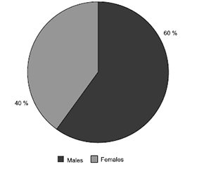
Are there neuromuscular junction disorders in the early stages of ALS? To answer this, we described 5 patients with ALS presented at Petre Sarajishvili Institute of Neurology in 2018–2022, 3 males and 2 females aged 50–61 years.
ALS diagnosis was based on clinical signs, the Gold Coast criteria, electromyography (Awaji), neuroimaging, blood and urine tests.

Results

We studied patients’ anamnesis, physical data, evaluated their mental, cognitive functions and neurological status. We have also interviewed family members, as it was often difficult for the patient to accurately describe the symptoms.
At the early stage of the disease, only asymmetric ptosis and diplopia were noted, which did not improve on pyri-dostigmine or steroids. Acetylcholine receptor antibodies were mildly positive only in one patient. Thymoma was excluded.
The neurophysiological study showed only marked neuromuscular transmission failure in orbicularis oculi, there were no clinical and electromyographic signs of motor neuron damage.
Approximately 2 years later, all five patients developed clinical and electromyographic signs of ALS. According to our research, the diagnosis of ALS is difficult until muscle atrophy and tremors are detected.
There are relatively less cases of ALS (2 patients) with symptoms on one or both legs. At this time, the patients felt uncomfortable while walking, the ankle lost its flexibility, its range of motion was limited. Muscle weakness is expressed; there were muscle spasms, increase of deep reflexes or expansion of the reflexogenic zone, pathological reflexes, pronounced muscle atrophy, increased spasticity. The upper limbs were less damaged, although the flexibility of the fingers is limited.

Discussion

Bulbar-onset ALS was detected in one patient with difficulty speaking, the patient spoke “through the nose”, later had difficulty swallowing. Speech disturbances (dysarthria, anarthria), voice production disorders (dysphonia, aphonia) were noted. Disappearance of gag reflex, salivation, breathing disorders were soon added to the symptoms. At the same time, in two patients with ALS confirmed by us, the symptoms included signs of both lower and upper motor neuron lesion. Damage to the upper motor neuron manifested itself in muscle hypertonia, hyperreflexia, pathological Babinski sign, and in case of the lower motor neuron lesion, muscle weakness, atrophy, involuntary fasciculations were present.

Conclusions

In the present study, neuromuscular junction disorders are found to play an important role in the pathogenesis of ALS and may serve as a useful early diagnostic marker.
 
Received 04.03.2024
Revised 15.03.2024
Accepted 24.03.2024

Список литературы

1. Kekenadze M, Rocca C, Kaiyrzhanov R, Nagy S, Kvirkvelia N, et al. Analysis of C9orf72 repeat expansions in Georgian patients with ALS. F1000Res. 2024 Mar 6;12:1113. doi: 10.12688/f1000research.138436.1.
2. Kekenadze M, et al. Clinical characteristics of ALS in Georgian patients. Georgian Medical News. 2021;319:71-75.
3. Paganoni S, Karam C, Joyce N, Bedlack R, Carter G. Comprehensive rehabilitative care across the spectrum of amyotrophic lateral sclerosis. NeuroRehabilitation. 2015;37(1):53-68. doi: 10.3233/NRE-151240.
4. Macpherson CE, Bassile CC. Pulmonary Physical Therapy Techniques to Enhance Survival in Amyotrophic Lateral Sclerosis: A Systematic Review. Journal of Neurologic Physical Therapy. 2016 Jul;40(3):165-175. doi: 10.1097/NPT.0000000000000136.
5. Moens J. Brain Implant Allows Fully Paralyzed Patient to Communicate. The New York Times. 2022 Mar 22.

Вернуться к номеру