Архів офтальмології та щелепно-лицевої хірургії України Том 1, №1, 2024
Вернуться к номеру
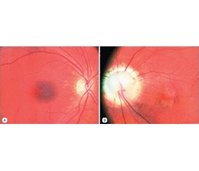
Клінічний випадок morning glory syndrome
Авторы: Венгер Л.В. (1), Коновалова Н.В. (1), Іваницька О.В. (1), Храменко Н.І. (2), Гузун О.В. (2), Журавок Ю.О. (1, 2)
(1) - Одеський національний медичний університет, м. Одеса, Україна
(2) - ДУ «Інститут очних хвороб і тканинної терапії імені В.П. Філатова НАМН України», м. Одеса, Україна
Рубрики: Офтальмология
Разделы: Справочник специалиста
Версия для печати
Актуальність. Рідкісна вроджена патологія, синдром в’юнка, або синдром ранкового сяйва (англ. morning glory syndrome), є лійкоподібною екскавацією заднього полюса очного яблука із залученням диска зорового нерва. Мета роботи: навести клінічний випадок рідкого захворювання та продемонструвати можливість лікування функціональних порушень у пацієнта з morning glory syndrome. Матеріали та методи. У наше дослідження ми включили випадок захворювання у чоловіка з монолатеральним синдромом morning glory. Проведено повне комплексне офтальмологічне обстеження. Результати. Аномалія morning glory являє собою вроджене лійкоподібне поглиблення перипапілярної сітківки та зорового нерва із залученням диска зорового нерва, яке пов’язане з аномаліями ока та головного мозку. Характерні зміни очного дна: збільшений, втягнутий диск зорового нерва з білою фіброгліальною тканиною в його центрі. Висновки. Morning glory syndrome — патологія, патогенез якої досі повністю не вивчений. Захворювання може діагностуватися і в зрілому віці, якщо пацієнти з різних причин не звертаються до офтальмолога.
Background. A rare congenital pathology, morning glory syndrome, is a funnel-shaped excavation of the posterior pole of the eyeball involving the optic disc. Aim: to present a clinical case of a rare disease and the possibility of treatment of functional disorders in a patient with morning glory syndrome. Materials and methods. In our study, we included a case of a man with unilateral morning glory syndrome. A full comprehensive ophthalmic examination was performed. Results. Morning glory anomaly is a congenital funnel-shaped deepening of the parapapillary retina and optic nerve involving the optic disc, which is associated with eye and brain anomalies and is characterized by a syndromic manifestation. Characteristic changes of the fundus: an enlarged, retracted disc of the optic nerve with white fibroglial tissue in its center. Conclusions. Morning glory syndrome is a rare congenital pathology whose pathogenesis is still not fully understood. The case described in this work indicates that the disease is often a one-sided pathology, which is detected in early childhood, but diagnosis also can be made in adulthood, when vision remains high, and patients do not consult an ophthalmologist.
morning glory syndrome; патологія диска зорового нерва; оптична когерентна томографія; флуоресцентна ангіографія; комп’ютерна томографія орбіти; ресвератрол
morning glory syndrome; optic disc abnormality; optical coherence tomography; fluorescein angiography; computed tomography of the orbit; resveratrol
Для ознакомления с полным содержанием статьи необходимо оформить подписку на журнал.
- Kindler P. Morning Glory Syndrome: Unusual Congenital Optic Disk Anomaly. Am J Ophthalmol. 1970;69(3):376-84. https://doi.org/–10.1016/0002-9394(70)92269-5.
- Handmann M. Erbliche vermutlich angeborene zentrale gliose Entartung des Sehnerven mit besonderer Beteilingung der Zen-tralgefaesse. Klin. Monatsbl. Augenheilkd. 1929;83:145.
- Reis W. Eine wenig bekannte typische Missbildung am Sehnerveneintritt: unschriebene Grubenbildung auf der Papilla n. optici. Z. Augenheilkd. 1908;19:505.
- Osaguona V.B., Momoh R.O. Morning Glory Syndrome in a Nigerian: A Case Report. Journal of the West African College of Surgeons. 2017;7:128-134.
- Jeng-Miller K.W., Cestari D.M., Gaier E.D. Congenital ano–malies of the optic disc: Insights from optical coherence tomography imaging. Curr. Opin. Ophthalmol. 2017;28:579-586. doi: 10.1097/ICU.0000000000000425;
- Cennamo G., de Crecchio G., Iaccarino G., Forte R., Cennamo G. Evaluation of morning glory syndrome with spectral optical coherence tomography and echography. Ophthalmology. 2010;117:1269-1273. doi: 10.1016/j.ophtha.2009.10.045.
- Cennamo G., Rossi C., Ruggiero P., de Crecchio G., Cennamo G. Study of the Radial Peripapillary Capillary Network in Congenital Optic Disc Anomalies with Optical Coherence Tomography Angiography. Am. J. Ophthalmol. 2017;176:1-8. doi: 10.1016/j.ajo.2016.12.016.
- Sevgi D.D., Orge F.H. Contractile morning glory disk anomaly: Analysis of the cyclic contractions and literature review. J. AAPOS. 2020;24:99.e1-99.e6. doi: 10.1016/j.jaapos.2020.01.009.
- Loudot C., Fogliarini C., Baeteman C., Mancini J., Girard N., Denis D. Rééducation de la part fonctionnelle de l’amblyopie dans un Morning Glory syndrome. Journal Français D’Ophtalmologie. 2007;30:998-1001. https://doi.org/10.1016/S0181-5512(07)79276-8.
- Dedhia C.J., Gogri P.Y., Rani P.K. Rare Bilateral Presentation of Morning Glory Disc Anomaly. BMJ Case Reports. 2016;2016: bcr2016215846. https://doi.org/10.1136/bcr-2016-215846.
- Kouassi F.X., Koman C.E., Diomandé I.A., Somahoro M., Sowagnon T.Y.C., Kra A.N.S., Koffi K.V. Morning Glory Syndrome: A Case Report. Journal of Ophthalmology. 2017;2:000129. https://doi.org/10.23880/OAJO-16000129.
- Napo Abdoulaye et Sidibe Mohamed Kolé. Le syndrome du soleil levant. Pan African Medical Journal. 2017;26:176. https://doi.org/10.11604/pamj.2017.26.176.11445.
- Gupta A., Singh P., Tripathy K. Morning Glory Syndrome. [Updated 2023 Aug 25]. In: StatPearls [Internet]. Treasure Island (FL): StatPearls Publishing; 2024 Jan-. Available from: https://www.ncbi.nlm.nih.gov/books/NBK580490.
- Ponnatapura J. Morning glory syndrome with Moyamoya di–sease: A rare association with role of imaging. Indian J Radiol Imaging. 2018 Apr-Jun;28(2):165-168. doi: 10.4103/ijri.IJRI_219_17. PMID: 30050238; PMCID: PMC6038211.
- Zou Y., She K., Hu Y., Ren J., Fei P., Xu Y., Peng J., Zhao P. Clinical and Echographic Features of Morning Glory Disc Anomaly in Children: A Retrospective Study of 249 Chinese Patients. Front Med (Lausanne). 2022 Jan 24;8:800623.
- Sawada Y., Fujiwara T., Yoshitomi T. Morning glory disc anomaly with contractile movements. Graefes Arch Clin Exp Ophthalmol. 2012;250:1693-5.
- Kumar J., Adenuga O.O., Singh K., Ahuja A.A., Kannan N.B., Ramasamy K. Clinical characteristics of morning glory disc anomaly in South India. Taiwan J Ophthalmol. 2020 Oct 19;11(1):57-63.
- Ceynowa D.J., Wickström R., Olsson M., Ek U., Eriksson U., Wiberg M.K., Fahnehjelm K.T. Morning glory disc anomaly in childhood — A population-based study. Acta Ophthalmol. 2015;93:626-634. doi: 10.1111/aos.12778. [PubMed] [CrossRef]
- Fattah M.A., Reginald Y.A. Morning Glory Disc Ano–maly: A Baby with Strabismus and an Abnormal Optic Disc. In: Heidary G., Phillips P.H. (eds) Fundamentals of Pediatric Neuro-–Ophthalmology. Springer, Cham, 2023. https://doi.org/–10.1007/ 978-3-031-16147-6.
- Zheng S., Cao J.F., Wang X.Y., Wu B., Wang Z.J., Xiao B., Duan J.L., Hu P. Multimodal imaging of morning glory syndrome with persistent hyperplastic primary vitreous. J. Clin. Ultrasound. 2023;51:1364-1365. doi: 10.1002/jcu.23562. [PubMed] CrossRef] [Google Scholar]
- Chen Y.N., Patel C.K., Kertes P.J., Devenyi R.G., Blaser S., Lam W.C. Retinal Detachment and Retrobulbar Cysts in a Large Cohort of Optic Nerve Coloboma. Retina. 2018;38:692-697. doi: 10.1097/IAE.0000000000001594.
- Traboulsi E.I. [et al.]. Aniridia, atypical iris defects, optic pit and the morning glory disc anomaly in a family. Genet. 1986;7: 131-135.
- Safari A. Morning glory syndrome associated with multiple sclerosis. Iran J. Neurol. 2014;13(3):177-180.
- Cennamo G., Rinaldi M., Concilio M., Costagliola C. Congenital Optic Disc Anomalies: Insights from Multimodal Imaging. J Clin Med. 2024 Mar 6;13(5):1509. doi: 10.3390/jcm13051509. PMID: 38592429; PMCID: PMC10932420.
- Ní Leidhin C., Erickson J.P., Bynevelt M., Lam G., Lock J.H., Wang G., et al. (What’s the story) morning glory? MRI findings in morning glory disc anomaly. Neuroradiology. 2024 May 8. doi: 10.1007/s00234-024-03375-2.
- Nguyen D.T., Boddaert N., Bremond-Gignac D., Robert M.P. Аномалії зорового нерва при аномалії диска іпомеї: дослідження МРТ. J Neuroophthalmol. 2022;42:199-202.
- She K., Zhang Q., Fei P., Peng J., Lyu J., Li Y., Huang Q., Zhao P. Peripheral Retinal Nonperfusion in Pediatric Patients with Morning Glory Syndrome. Ophthalmic Surg. Lasers Imaging Retin. 2018;49:674-679.
- Duvall J., Miller S.L., Cheatle E., Tso M.O. Histopathologic study of ocular changes in a syndrome of multiple congenital anomalies. Am. J. Ophthalmol. 1987;103:701-705.
- Thuma T.B.T., Procopio R.A., Jimenez H.J., Gunton K.B., Pulido J.S. Hypomorphic variants in inherited retinal and ocular diseases: A review of the literature with clinical cases. Surv Ophthalmol. 2024 May-Jun;69(3):337-348. doi: 10.1016/j.survophthal.2023.11.006. Epub 2023 Nov 29. PMID: 38036193.
- Delmas D., Solary E., Latruffe N. Resveratrol, a phytochemi–cal inducer of multiple cell death pathways: Apoptosis, autophagy and mitotic catastrophe. Curr. Med. Chem. 2011;18:1100-1121. doi: 10.2174/092986711795029708.

