Журнал «Боль. Суставы. Позвоночник» Том 14, №3, 2024
Вернуться к номеру
Клініко-морфометричні зміни шийного відділу хребта в людей і тварин з болем у шиї
Авторы: Андреєва Т.О. (1), Стоянов О.М. (2), Мірджураєв Е.М. (3), Чеботарьова Г.М. (4), Калашніков В.Й. (5), Вастьянов Р.С. (2), Дарій В.І. (6)
(1) - Чорноморський національний університет імені П. Могили МОН України, м. Миколаїв, Україна
(2) - Одеський національний медичний університет МОЗ України, м. Одеса, Україна
(3) - Ташкентський інститут удосконалення лікарів, м. Ташкент, Узбекистан
(4) - Одеський національний політехнічний університет МОН України, м. Одеса, Україна
(5) - Харківський національний медичний університет МОЗ України, м. Харків, Україна
(6) - Запорізький державний медико-фармацевтичний університет МОЗ України, м. Запоріжжя, Україна
Рубрики: Ревматология, Травматология и ортопедия
Разделы: Клинические исследования
Версия для печати
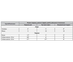
Актуальність. Відома висока активність, рухливість шиї, її кістково-хрящового, м’язового апарату тощо. При цьому дегенеративно-дистрофічні процеси у шийному відділі хребта є нагальною проблемою. Метою дослідження було визначити клініко-морфометричні зміни шийного відділу хребта у людей і тварин з болем у шиї на ґрунті клініко-неврологічного обстеження, визначення щільності тіл хребців, їх конфігурації та співвідношення задля своєчасної корекції, прогнозу цієї патології. Матеріали та методи. У людей і тварин вивчали інтенсивність болю за допомогою адаптованих ВАШ, комп’ютерно-томографічні показники з вимірюванням щільності тіл хребців, морфометричних показників з акцентом локалізації на рівні С5–С7. Усі дослідження проводилися відповідно до сучасних біоетичних стандартів. Результати. Патологія конфігурації хребта зареєстрована у 84,6 % обстежених у вигляді кутового кіфозу або випрямленого лордозу, частіше у жінок. У собак і котів зміни спостерігали в 34,7 % випадків. Нормальна конфігурація частіше: у кішок — 78,6 % і собак дрібних порід — 78,5 %, у великих порід собак — лише у 26,3 %, при цьому деформації зустрічалися частіше, ніж у кішок і дрібних собак (у 2,7 раза і більше). Щільність тіл хребців вздовж хребта в усіх групах зменшувалася в каудальному напрямку, у людей ця відмінність становила 18,1 %. У кішок така відмінність становила в середньому 2,7 %, у собак дрібних порід вона була значнішою — 7,5 %, а у великих порід досягала 14,3 %. Максимальна відповідність змін відмічена в людей і собак великих порід. Висновки. Тварини, особливо собаки великих порід, можуть бути моделлю для вивчення етіопатогенетичних чинників, перебігу та прогнозу дегенерації кістково-хрящового апарату шийного відділу хребта.
Background. The neck, its bone-cartilaginous and muscle apparatus, etc. high activity is well known. Cervical spine degenerative-dystrophic processes are considered to be an urgent problem. The purpose of the study was to determine the clinical and morphometric changes of the cervical spine in humans and animals with neck pain based on a clinical and neurological examination, determination of the vertebral body density, their configuration, and ratio for timely correction and prognosis of this pathology. Materials and methods. Pain intensity was studied in humans and animals using the adapted visual-analogue scale. The indexes of computer tomography with measurement of vertebral body density, and morphometric indexes with an emphasis on C5-C7 level were also studied. All studies were conducted following existing bioethical standards. Results. The pathology of spine configuration was registered in 84.6 % of the examined patients in the form of angular kyphosis or straightened lordosis, more often in women. It was observed in 34.7 % of cases in dogs and cats. The normal configuration is more common: in cats — 78.6 % and in dogs of small breeds — 78.5 %, in large breed dogs — only 26.3 %, and deformations were more frequent than in cats and small dogs (2.7 times more). The cervical vertebrae bodies density in all groups decreased toward the caudal direction with a difference of 18.1 % in humans. In cats — 2.7 %, in dogs of small breeds, it was higher (7.5 %), and in large breed dogs, it reached 14.3 %. The maximum deviations of the studied indicators were found in humans and maximally coincided with those in dogs of large breeds. Conclusions. Thus, animals, especially dogs of large breeds, can serve as a model for studying etiopathogenetic factors, the course, prognosis of degeneration of the bone-cartilage apparatus.
дегенеративно-дистрофічна патологія хребта; шийний відділ; люди; тварини; біль; ішемія; неврологічні розлади; комп’ютерна томографія; денситометрія
spine degenerative-dystrophic pathology; cervical region; people; animals; pain; ischemia; neurological disorders; computer tomography; densitometry
Для ознакомления с полным содержанием статьи необходимо оформить подписку на журнал.
- Kaiser JT, Reddy V, Launico MV, Lugo-Pico JG. Anatomy, Head and Neck: Cervical Vertebrae. [Updated 2022 Oct 6]. In: StatPearls [Internet]. Treasure Island (FL): StatPearls Publishing; 2023 Jan. Available from: https://www.ncbi.nlm.nih.gov/books/NBK539734/.
- Charbonneau L, Watanabe K, Chaalala C, Bojanowski MW, Lavigne P, Labidi M. Anatomy of the craniocervical ujnction — A review. Neurochirurgie. 2024 May;70(3):101511. doi: 10.1016/j.neuchi.2023.101511.
- Andreeva T, Chebotaryova G, Stoyanov O, Vastyanov R, Kalashnikov V, StoyanovA. Acquired stenosis of the spinal canal. A comparative study of humans and dogs. International neurological journal. 2022;18(4):24-29. Ukrainian. doi: 10.22141/2224-0713.18.4.2022.955.
- Diseases and Injuries Collaborators. Global burden of 369 diseases and injuries in 204 countries and territories, 1990–2019: a systematic analysis for the Global Burden of Disease Study 2019. Lancet. 2020 Oct;396(10258):1204-1222. doi: 10.1016/S0140-6736(20)30925-9.
- Saini A, Mukhdomi T. Cervical Discogenic Syndrome. [Updated 2022 Apr 5]. In: StatPearls [Internet]. Treasure Island (FL): StatPearls Publishing; 2022 Jan. Available from: https://www.ncbi.nlm.nih.gov/books/NBK555960.
- Іпатов А.В., Мороз О.М., Гондуленко Н.О. та ін. Основні показники інвалідності та діяльності медико-соціальних експертних комісій України за 2018 рік: аналітико-інформаційний довідник. За ред. Р.Я. Перепеличної. Дніпро: Акцент ПП, 2019. 180 с.
- Doskaliuk B, Zaiats L, Yatsyshyn R, Gerych P, Cherniuk N, Zimba O. Pulmonary involvement in systemic sclerosis: exploring cellular, genetic and epigenetic mechanisms. Rheumatol Int. 2020 Oct;40(10):1555-1569. doi: 10.1007/s00296-020-04658-6.
- Dolhopolov OV, Polishko VP, Yarova ML. Epidemiology of Diseases of the Musculoskeletal System in Ukraine for the Period 1993-2017. Herald of orthopedics, traumatology and prosthetics. 2019;4:101-108. Ukrainian. doi: 10.37647/0132-2486-2019-103-4-96-104.
- GBD 2017 Disease and Injury Incidence and Prevalence Collaborators. Global, regional, and national incidence, prevalence, and years lived with disability for 354 diseases and injuries for 195 countries and territories, 1990–2017: a systematic analysis for the Global Burden of Disease Study 2017. Lancet. 2018 Nov 10;392(10159):1789-1858. doi: 10.1016/S0140-6736(18)32279-7.
- Lacroix M, Nguyen C, Burns R, Laporte A, Rannou R, Feydy A. Degenerative Lumbar Spine Disease: Imaging and Biomechanics. Semin Musculoskelet Radiol 2022 Aug;26(4):424-438. doi: 10.1055/s-0042-1748912.
- Brinjikji W, Luetmer PH, Comstock B, Bresnahan BW, Chen LE, Deyo RA. et al. Systematic Literature Review of Imaging Features of Spinal Degeneration in –Asymptomatic Populations. AJNR Am J Neuroradiol. 2015 Apr;36(4):811-816. doi: 10.3174/ajnr.A4173.
- Been E, Shefi S, Soudack M. Cervical lordosis: the effect of age and gender. Spine J. 2017 Jun;17(6):880-888. doi: 10.1016/j.spinee.2017.02.007.
- Baba H, Uchida K, Maezawa Y, Furusawa N, Azuchi M, Imura S. Lordotic alignment and posterior migration of the spinal cord following en bloc open-door laminoplasty for cervical myelopathy: a magnetic resonance imaging study. J Neurol. 1996;85:626-32.
- Guo GM, Li J, Diao QX, Zhu TH, Song ZX, Guo YY, Gao YZ. Cervical lordosis in asymptomatic individuals: a meta-analysis. J Orthop Surg Res. 2018;13(1):147. doi: 10.1186/s13018-018-0854-6.
- Highsmith JM, Dhall SS, Haid RW, Rodts GE, Mummaneni PV. Treatment of cervical stenotic myelopathy: a cost and outcome comparison of laminoplasty versus laminectomy and lateral mass fusion. J Neurosurg Spine. 2011 May;14(5):619-625. doi: 10.3171/2011.1.SPINE10206.
- Swank ML, Sutterlin CE, Bossons CR, Dials BE. Rigid internal fixation with lateral mass plates in multilevel anterior and posterior reconstruction of the cervical spine. Spine. 1997;22:274-82.
- Wang Z, Luo G, Yu H, Zhao H, Li T, Yang H, Sun T. Comparison of discover cervical disc arthroplasty and anterior cervical discectomy and fusion for the treatment of cervical degenerative disc diseases: A meta-analysis of prospective, randomized controlled trials. Front Surg. 2023;10:1124423. doi: 10.3389/fsurg.2023.1124423.
- Калашников В.Я., Стоянов О.М., Вастьянов Р.С., Калашнікова І.В., Бакуменко І.К. Особливості мозкового кровообігу у хворих з різними видами головного болю. Науковий вісник Ужгородського університету. Серія «Медицина». 2023. 1(67). С. 115-120.
- Patel PD, Arutyunyan G, Plusch K, Vaccaro JrA, Vaccaro AR. A review of cervical spine alignment in the normal and degenerative spine. J Spine Surg. 2020 Mar;6(1):106-123. doi: 10.21037/jss.2020.01.10.
- Wang T, Ma J, Hogan AN, Fong S, Licon K, Tsui B. et al. Quantitative translation of dog-to-human aging by conserved remodeling of the DNA methylome. Cell Syst. 2020 Aug 26;11(2):176-185.e6. doi: 10.1016/j.cels.2020.06.006.
- Wysocki MA, Feranec RS, Tseng ZJ, Bjornsson CS. Using a Novel Absolute Ontogenetic Age Determination Technique to Calculate the Timing of Tooth Eruption in the Saber-Toothed Cat, Smilodon fatalis. PLoS One. 2015 Jul 1;10(7):e0129847. doi: 10.1371/journal.pone.0129847.
- Стоянов О.М., Вастьянов Р.С., Скоробреха В.З. Патофізіологічні механізми нейровегетології болю. Одеса: Астро-Принт, 2015. 112 с.
- Hielm-Björkman AK, Kapatkin AS, Rita HJ. Reliability and validity of a visual analogue scale used by owners to measure chronic pain attributable to osteoarthritis in their dogs. Am J Vet Res. 2011 May;72(5):601-607. doi: 10.2460/ajvr.72.5.601.
- Mathews K, Kronen PW, Lascelles D, Nolan A, Robertson S, Steagall PV. et al. Guidelines for recognition, assessment and treatment of pain. J Small Anim Pract. 2014 Jun;55(6):10-68. doi: 10.1111/jsap.12200.
- Morales-Avalos R, Leyva-Villegas J, Sánchez-Mejorada G, Cárdenas-Serna M, Vílchez-Cavazos F, Martínez-Ponce De León A. et al. Age- and sex-related variations in morphometric characteristics of thoracic spine pedicle: A study of 4,800 pedicles. Clin Anat. 2014 Apr;27(3):441-450. doi: 10.1002/ca.22359.
- Tjahjadi D, Onibala MZ. Torg ratios based on cervical lateral plain films in normal subjects. Universal Medicine. 2010;29(1):8-13. doi: 10.18051/UnivMed.2010.v29.8-13.
- Tao Y, Niemeyer F, Galbusera F, Jonas R, Samart–zis D, Vogele D. et al. Sagittal wedging of intervertebral discs and vertebral bodies in the cervical spine and their associations with age, sex and cervical lordosis: A large-scale morphological study. Clin Anat. 2021 Oct;34(7):1111-1120. doi: 10.1002/ca.23769.
- Patel EA, Perloff MD. Radicular Pain Syndromes: Cervical, Lumbar, and Spinal Stenosis. Semin Neurol. 2018 Dec;38(6):634-639. doi: 10.1055/s-0038-1673680.
- Andreyeva TO, Stoyanov OM, Chebotaryova GM, Vastyanov RS, Kalashnikov VI, Stoyanov AO. Comparative clinical and morphometric investigations of cervical stenosis of the spinal canal in humans and dogs. Regulatory Mechanisms in Biosystems. 2022;13(3):301-307. doi: 10.15421/022239.
- Вастьянов Р.С., Стоянов О.М., Бакуменко І.К. Системна патологічна дезінтеграція при хронічній церебральній ішемії. Експериментально-клінічні аспекти. Sa–arbrucken: LAP Lambert Academic Publishing, 2015. 169 с.
- Fakhoury J, Dowling TJ. Cervical Degenerative Disc Disease. [Updated 2023 Aug 14]. In: StatPearls [Internet]. Treasure Island (FL): StatPearls Publishing; 2024 Jan. Available from: https://www.ncbi.nlm.nih.gov/books/NBK560772.
- Stoyanov AN, Kalashnikov VI, Vastyanov RS, Pulyk AR, Son AS, Kolesnik OO. State of autono–mic regulation and cerebrovascular reactivity in patients with headache with arterial hypertension. Wiadomości Lekarskie. 2022;75(9p2):2233-2237. doi. 10.36740/WLek202209210.
- Dobran M, Nasi D, Benigni R, Colasanti R, Gladi M, Iacoangeli M. Cervical lordosis after subaxial spinal trauma surgery: relationship with neck pain and stiffness. G Chir. 2019 Nov-Dec;40(6):513-519.
- Grob D, Frauenfelder H, Mannion AF. The association between cervical spine curvature and neck pain. Eur Spine J. 2007 May;16(5):669-678. doi: 10.1007/s00586-006-0254-1.

