
Журнал «Здоровье ребенка» 6 (57) 2014
Вернуться к номеру
Cardiac arrhythmias: atrial flutter of newborn the case from practice
Авторы: Klimenko Т.M., Каrаpеtyan О.Yu., Zakrevskiy А.N., Serdseva Е.А. - Kharkov Medical Academy of Postgraduate Education; Karatay О.S., Soloshenko I.V., Melnichuk О.P. - Kharkov City Perinatal Center, Ukraine
Рубрики: Педиатрия/Неонатология
Разделы: Справочник специалиста
Версия для печати
Atrial flutter (AF) - a kind of atrial fibrillation, in which there is a rapid rhythmic excitation and contraction of the atria with a frequency of 350-400-480 pulses/min [1]. AF is caused by activation of ectopic pacemaker, located in the atria, it becomes a pacemaker, replacing work sinus [2]. In pediatric practice, atrial flutter is quite rare, accounting for about 2% of all fetal arrhythmias, and 20-25% of all perinatal persistent tachycardia. Cause of AF are organic heart disease with the localization of the pathological process in the atria: congenital heart disease, cardiomyopathy, fibroelastosis, carditis [3]. However, 14-30% of AF occurs in the absence of organic lesions of the myocardium and is regarded as idiopathic [3, 5].
Higher rates of complications in the form of heart failure, thromboembolism, arrhythmogenic cardiomyopathy, heart failure, leading to high (17-21 %) mortality, pose the problem of diagnosis and treatment of neonatal AF in some of the most pressing pediatric problems [6].
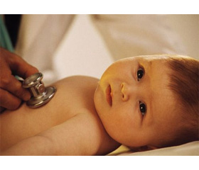
Clinically characterized by concern for the newborn child, possible refusal to eat, vomiting, quickly develop symptoms of heart failure. Typical auscultatory signs AF no. The diagnosis is established by using an electrocardiogram (ECG). Direct mechanisms, leading to a very high frequency excitation and reduction in their flutter are increasing automaticity cells in atrial conduction system or mechanism of «re-entry». As a result of frequent rhythmic atrial excitation on the ECG recorded frequent regular atrial wave, denoted F (flutter), having the characteristic sawtooth shape [6 , 7]. Ventricular QRS complexes with AF have the usual form, as excitation passes to the ventricles in the usual way . The frequency of ventricular contraction in AF usually less frequency atrial contraction due to the presence of functional AV block: 2:1, or 3:1, or 4:1.
Treatment of AF involves three steps: conversion rate, maintenance of sinus rhythm and control of atrioventricular conduction and ventricular rhythm. According to the requirements of the order of Ministry of Health of Ukraine № 362 dated 19.07.2005, the drugs of choice for the relief of AF are digoxin and amiodarone [8]. With recurrent forms of AF, no effect of drug cardioversion or contraindications for its use, the method of choice to restore the heart rhythm is electrical cardioversion single discharge rate of 5.5 J/kg [6, 9].
Give their own observation of the patient with atrial flutter.
Patient, 24 years old woman was admitted to Kharkov City Perinatal Center due to the presence of fetal tachycardia with a heart rate of 170 in 1 minute, which was interpreted as a manifestation of fetal distress. From history we know that this second pregnancy (the first ended with the birth of a healthy baby), gestational age of 36 weeks, proceeded physiological, factors burdening family history is not revealed. During fetal ultrasound with Doppler velocimetry revealed uncoordinated contraction of the heart chambers: atrium with reduced frequency of more than 200 in 1 minute and the ventricles - 170 in 1 minute. Produced delivery by cesarean section. Child (a girl ) was born with a mass 2490.0 , size 45-32-33 cm in the state Apgar 5-7 points. The baby's condition at birth is regarded as serious by cardiorespiratory failure requiring mechanical ventilation prosthetics. Since the birth of a child in need of mechanical ventilation, there was tachycardia with a heart rate of 190-220 in 1 minute. X-ray examination of the chest - Moderate interstitial pulmonary edema, a typical position of the heart shape-mitral, contours-clear, cardiothoracic index - 55 %. Holter ECG monitoring revealed atrial flutter.
For medication of the cardioversion assigned amiodarone, 5 mg/kg intravenously over 6 hours. Heart rhythm to normal after 5.5 hours from the start of infusion. The child's condition improved, and 6 hours after successful cardioversion transferred to spontaneous breathing and started enteral feeding. Amiodarone is continued at a dose of 10 → 5 mg/ kg /day, with the transition to the tablet form of application.
After the drug cardioversion held ultrasonic diagnostic cardiac Doppler mapping. Conclusion: Contractile force saved. Hypokinetic type of the central hemodynamics: ejection fraction - 56.94%, functioning fetal communication: the foramen ovale 3.6 mm, left-right rejection, the ductus arteriosus is not hemodynamically significant. Diastolic dysfunction of the right ventricle was diagnosed.
Child was discharged from hospital at the age of 12 days of life in the presence of regular sinus rhythm to monitor the cardiologist.
1. The presence of a newborn child suspected pathology of the cardiovascular system is necessary to conduct echocardiography with Doppler mapping, ECG, possibly with Holter monitoring.
2. Method of choice in newly diagnosed atrial flutter in newborns is a drug cardioversion using class III antiarrhythmic drug amiodarone. Conducting Holter ECG monitoring in parallel drug cardioversion to evaluate the effectiveness of interventions in real time.
3. Conduct electrical cardioversion newborn advisable for recurrent forms of atrial flutter, the absence of effect of drug cardioversion or contraindications for its use.