
Журнал «Здоровье ребенка» 4 (64) 2015
Вернуться к номеру
Immunologic reactivity in adolescents with duodenal ulcer
Авторы: Parkhomenko L.K., Strashok L.A., Zavelya E.M., Isakova M.Yu., Eshenko A.V. - Kharkiv Medical Academy of Post-graduate Education; Evdokimova T.V. - SI «Institute of Children and Adolescents Health Care of the NAMS of Ukraine»
Рубрики: Педиатрия/Неонатология
Разделы: Справочник специалиста
Версия для печати
According to WHO, 10-15% of the population of the developed countries suffer from peptic ulcer. Still the prevalence of peptic ulcer does not have a tendency to decrease in Ukraine, and occurring complications are often dangerous to the patient's life and require surgical correction. Our research shows that peptic ulcer may occur in the teenage years. Considering that peptic ulcer is a chronic relapsing disease, which tends to progression, active role of immune changes in the course of the disease is confirmed in our time. It is known that prevalence of diseases of the digestive system is increased in adolescence. This period is critical for violation of immunological reactivity too.
The aim of our study was to examine the state of non-specific and specific protective factors in adolescents with duodenal ulcer.
The study included 235 adolescents with duodenal ulcer (136 boys and 99 girls). Immunologic reactivity was assessed by standard techniques. The level of cytokines - IL-1β, IL-4, IL-6 and TNF-α were determined by enzyme-linked immunosorbent assay (ELISA) in serum. Using morphological and immunohistochemical methods according to standard procedures we investigated mucous coat of stomach and duodenum, cytokine-producing cells were differentiated using monoclonal antibodies to IL-1β, IL-4, IL-6, TNF-α.
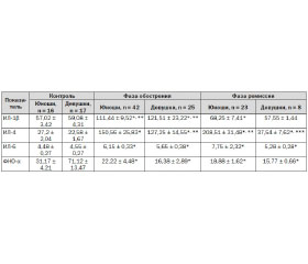
Results and discussion. Study revealed inhibition of cell-mediated immunity and tension of antibody-mediated immunity in adolescents with erosive and ulcerative lesions of the stomach and duodenum in acute period. It was revealed by a significant reduction of the absolute and relative number of the T-lymphocytes, immunoregulatory index and by the decreasing tendency of the absolute and relative number of B-lymphocytes despite higher levels of IgA and circulating immune complexes (CIC). Immunological imbalance in adolescents with duodenal ulcer contributed to chronicity of the process. That exhausted body adaptive systems and led to the secondary immune deficiency. Significantly increased levels of IL-1β, IL-6 (1.5 - 2 times) and IL-4 (5 times) proinflammatory cytokines were revealed in blood of all adolescents with duodenal ulcer in the acute phase of the disease. The concentration of TNF-α in blood was decreased by 1.5 times for boys and by 5 times for girls. Except for TNF-α, the concentration of cytokines was decreased to benchmarks as for girls, in the remission phase of the disease. There was only a decreasing tendency of IL-1β and IL-6, the concentration of IL-4, on the contrary, was significantly increased for boys. This fact indicated a more favorable course of the duodenal ulcer among girls; increasing the concentration of IL-4 , apparently, contributed to the oppression of cell-mediated immunity and potentiated anaphylactic reactions in the mucous coat in the remission phase in boys. Perhaps this explains the progressive course of destructive diseases of the upper gastrointestinal tract in males.
Immunohistochemical examination of mucous coat of stomach and duodenum revealed increasing cytokine-producing cells infiltration. Study showed weak TNF-α producing cells infiltration of mucous coat in adolescents with duodenal ulcer, unlike most studies show. This fact contributes to the low level of apoptosis and potentiates the proliferative processes. We used analysis of paired correlation coefficients between the concentration of cytokines in the blood and immunohistochemical studies. IL-1β and IL-6 potentiate the height of the mucous coat of the gastric and duodenum and the IL-1β, IL-6 producing cells infiltration. Anti-inflammatory effect of IL-4 was manifested by the presence of a negative correlation with the number of IL-1β and IL-6 producing cells in the mucous coat.
Conclusions:
1. Duodenal ulcer is formed and proceeds on the background of oppression of cell-mediated immunity, tension of antibody-mediated immunity and cytokine imbalance in adolescence, which reflects disturbances in the regulation of immune response.
2. The level of cytokines in serum correlates with the level of cytokine-producing cells infiltration of mucous coat of stomach and duodenum.
3. General immunologic disorders and local inflammatory changes in the mucous coat of stomach and duodenum which were identified in adolescents with duodenal ulcer should be considered when carrying out rehabilitation.
1. Babaeva A.R., Rodionova O.N., Vidiker R.V. Tsitokinovaya regulyatsiya funktsional'nykh zabolevaniy zheludochno-kishechnogo trakta. Vestnik novykh meditsinskikh tekhnologiy. 2011;18(1):163-4. Russian.
2. Beylina N.I. Optimizatsiya okazaniya meditsinskoy pomoshchi obuchayushcheysya molodezhi s erozivno-yazvennymi zabolevaniyami gastroduodenal'noy zony v usloviyakh gorodskoy studencheskoy polikliniki [Extended abstract of candidate’s thesis]. Kazan, 2013. Russian.
3. Il'intseva N.V. Gumoral'nyy i sekretornyy immunnyy otvet k Helicobacter pylori u detey s khroonicheskimi vospalitel'nymi zabolevaniyami zheludka i dvenadtsatiperstnoy kishki [Extended abstract of candidate’s thesis]. Moskva, 2011. Russian.
4. Kuramshina O.A. Kliniko-patogeneticheskie osobennosti formirovaniya i techeniya yazvennoy bolezni dvenadtsatiperstnoy kishki u lits molodogo vozrasta [Extended abstract of Doctor’s thesis ]. Ufa, 2014. Russian.
5. Lebedev N.V., Klimov A.E. Yazvennye gastroduodenal'nye krovotecheniya. Moskva: BINOM; 2010. Russian.
6. Lobankov V. M. Yazvennaya bolezn': kontseptsiya populyatsionnoy tyazhesti. Vestnik khirurgicheskoy gastroenterologii. 2009;(1):19-25. Russian.
7. Reshetnikov O.V., Kurilovich S.A., Varaksin N.A. et al. Uroven' syvorotochnykh tsitokinov detey, infitsirovannykh razlichnymi shtammami Helicobacter pylori. Eksperimetal'naya i klinicheskaya gastroenterologiya. 2010;(9):52-4. Russian.
8. Tsaregorodtseva T.M., Serova T.I. Tsitokiny v gastroenterologii. Moskva; 2003. Russian.
9. Tsimmerman, Ya.S. Problema etiologii i patogeneza yazvennoy bolezni: perechityvaya V.Kh. Vasilenko. Klinicheskaya meditsina. 2011;(1):14-9. Russian.
10. Bardhan K.D, Royston C. Time, change and peptic ulcer disease in Rotherham, UK. Dig Liver Dis. 2008 Jul;40(7):540-6. doi: 1016/j.dld.2008.02.024.
11. David J. Bjorkman. Prevalence of Idiopathic PUD in a Developed Country. Aliment Pharmacol Ther. 2013 Oct;38:946.
12. Karen van Rensburg. Acid suppressants and peptic ulcer disease. SAPJ. 2010 Apr:33-7.
13. H.R. Rahimi, M. Rasouli, A. Jamshidzadeh, et al. New immunological investigations on Helicobacter pylori-induced gastric ulcer in patients. Microbiol Immunol. 2013 Jun;57(6):455-62. doi: 10.1111/1348-0421.12056.
14. M.H.Moller, S.Adamsen, M. Wojdemann, A.M. Moller. Perforated peptic ulcer: how to improve outcome? Scand J Gastroenterol. 2009;44(1):15-22. doi: 10.1080/00365520802307997.
15. Wang Y. R., Richter J. E, Dempsey D. T. Trends and outcomes of hospitalizations for peptic ulcer disease in the United States, 1993 to 2006. Ann Surg. 2010 Jan;251(1):51-8. doi: 10.1097/SLA.0b013e3181b975b8.