Журнал «Травма» Том 18, №3, 2017
Вернуться к номеру
Клінічні аспекти застосування фіксатора типу «ендобатон» для лікування травм і деформацій опорно-рухового апарату в умовах Обласної лікарні інтенсивного лікування м. Маріуполя
Авторы: Ставицький О.Б.(1), Пастернак Д.В.(1,2), Карпушкін О.В.(1), Абрамович Е.О.(1), Ямковий І.А.(1), Позняк О.С.(1)
(1) — Обласна лікарня інтенсивного лікування, м. Маріуполь, Україна
(2) — Донецький національний медичний університет, м. Лиман, Україна
Рубрики: Травматология и ортопедия
Разделы: Клинические исследования
Версия для печати
Проведено аналіз оперативного лікування 56 пацієнтів із пошкодженнями капсульно-зв’язкового апарату, сухожилків, переломами кісток та набутими деформаціями з використанням фіксації типу «ендобатон». Після оперативного втручання результати лікування спостерігалися в усіх пацієнтів у терміни від 4 до 36 місяців. Методика фіксації є доступною, економічно обґрунтованою, відносно простою в технічному виконанні з можливістю надійної фіксації пошкоджених структур та ранньою активізацією й реабілітацією хворого.
Проведен анализ оперативного лечения 56 пациентов с повреждениями капсульно-связочного аппарата, сухожилий, переломами костей и приобретенными деформациями с использованием фиксации типа «эндобаттон». После оперативного вмешательства наблюдение за результатами лечения проводилось у всех пациентов в сроки от 4 до 36 месяцев. Методика фиксации зарекомендовала себя как доступная, экономически обоснованная, относительно простая в техническом выполнении с возможностью надежной фиксации поврежденных структур и ранней активизацией и реабилитацией больного.
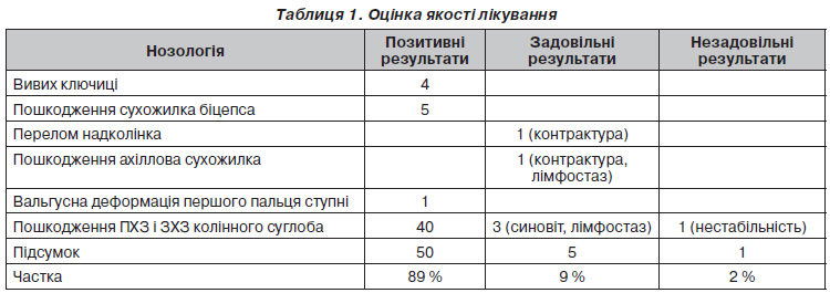
Background. At present, the damage to the capsular ligaments, joints of limbs are common injuries in the structure of the musculoskeletal trauma. Such injuries, as cruciate ligament rupture, dislocation of the clavicle, separation tendons (damage to the biceps tendon and Achilles tendon), in young people often result from direct effects (shock, falls) or rotational component due to the impact force and is accompanied by significant damage to the capsular ligament structures of the joint. In the elderly, against the background of degenerative-dystrophic changes in the bones and tendons, such injuries result from minor impact strength (sudden movement, lifting weights, daily work). Deformation related to the failure of tendon-ligament apparatus — transverse platypodia and valgus deformity of hallux of the foot. At present, one of the effective technologies of surgical treatment that meets modern requirements is considered to be endobutton fixation, which is used in surgical treatment of damages to the capsular ligaments, joints, tendon ruptures and their consequences, as well as acquired foot deformities. This gives an opportunity of quality repair of the damaged anatomical structures, providing stable fixation and the possibility of early activation and rehabilitation of the patient, which will restore the function of the injured extremity in small terms with minimal loss of quality of life. The purpose of the research is to assess the effectiveness of the endobutton fixation device in the surgical treatment of patients with damage to the capsular ligaments, joints and acquired foot deformities. Materials and methods. From 2014 to 2017, in the trauma department of Mariupol municipal hospital N 2, there were treated 56 patients with injury of the tendon-ligamentous apparatus, in whom the method of choice was to use endobutton fixation device: clavicle dislocation, dislocation fracture of acromial end of the clavicle, damage to the distal biceps tendon, proximal head biceps injury, Achilles tendon damage, bone exostosis of the second instep bone with valgus deformity, patella fracture, the rear cruciate ligament damage, anterior cruciate ligament damage. We have used the endobutton fixation device of three manufacturers: Endoprotec — in 19 patients, DePuy — in 15 patients, Smith & Nephew — in 22 patients. For navigation and minimally invasive endobutton introduction and fixation of the injuries, we have used arthroscopic navigation set for the plasty of cruciate ligament, cannulated drill manufactured by DePuy and Smith & Nephew companies. The average age of patients was 42 years (16–72 years). Of them, 16 persons were women and 40 — men. In 56 cases, the cause of injury was home injuries (falls, weight lifting, sports), in 1 case — work injury (in an accident), in 1 case — the disease. Results. Evaluation of the upper limb was performed on the DASH (The Disabilities of the Arm, Shoulder and Hand) scale. Additional assessment of the effectiveness of treating patients was conducted using visual analogue scale formed on the basis of the following criteria: scale of pain, limitation of movement in the joints of the limbs, deformation of the damaged zone, malnutrition, stability, ultrasound data, X-ray data, neurotrophic and neurological disorders, infectious complications. Positive results were considered to be: complete union and recovery of movement in the joints of the extremities, absence of muscle atrophy, neurotrophic and infectious complications. Satisfactory results were: damage coalescence, minor limitation of movement in the joints, hypotrophy, preservation of post-traumatic edema. The result was considered unsatisfactory, if there was no evidence of fracture union, instability persisted, joint contractures remained, there were purulent-septic and neurological complications, the need for reoperation. Conclusions. The fixation method is available, economically feasible, relatively simple in terms of technical performance, with the possibility of reliable fixation of damaged structures and early activation and rehabilitation of the patient. The use of navigation system and cannulated drills allows minimizing intraoperative trauma and increasing the accuracy of the choice of fixation sites, reducing the risk of osteoarthritis in damaged joints. The possibility of early functional load and isometric exercises in the postoperative period minimizes contracture of joints, reduces the overall rehabilitation period. A good cosmetic effect is achieved by minimizing the need for surgical approach and no need to remove the fixators.
хрестоподібні зв’язки; ахілловий сухожилок; вивих ключиці; перелом надколінка; плосковальгусна деформація стоп; оперативне лікування; «ендобатон»
крестообразные связки; ахиллово сухожилие; вывих ключицы; перелом надколенника; плосковальгусная деформация стоп; оперативное лечение; «эндобаттон»
cruciate ligaments; Achilles tendon; dislocation of the clavicle; patellar fracture; planovalgus deformity; surgical treatment; endobutton
Вступ
Матеріали та методи

Результати
Обговорення
Висновки
1. Травматология и ортопедия: Руководство для врачей: В 3 т. — Т. 2 / Под ред. Ю.Г. Шапошникова. — М.: Медицина, 1997. — 592 с.
2. Справочник травматолога / Под ред. Корж Н.А. — К.: ТОВ «Доктор-Медиа», 2009. — 504 с.
3. Травматология и ортопедия: Учебник / Х.А. Мусалатов, Г.С. Юмашев, Л.Л. Силин и др.; под ред. Х.А. Мусалатова, Г.С. Юмашева. — 4-е изд., перераб. и доп. — М.: Медицина, 1995. — 560 с.
4. Endobutton technique for the treatment of acute acromioclavicular joint / Özden R., Uruç V., Duman İ.G. [et al.] // Dicle Medical Journal. — 2014. — Vol. 41, № 2 — Р. 268-271.
5. Repair of the torn distal biceps tendon by endobutton fixation / Gupta R.K., Bither N., Singh H. [et al.] // Indian J. Orthop. — 2012. — Vol. 46, № 1. — P. 71-76.
6. Acromioclavicular and coracoclavicular cerclage reconstruction for acute acromioclavicular joint dislocations / Lädermann A., Grosclaude M., Lübbeke A. [et al.] // J. Shoulder Elbow Surg. — 2011. — Vol. 20, № 3. — P. 401-408.
7. Mid to long-term results of open acromioclavicular joint reconstruction using polydioxansulfate cerclage augmentation / Greiner S., Braunsdorf J., Perka C. [et al.] // Arch. Orthop. Trauma Surg. — 2009. — № 129. — P. 735-740.
8. Prodromos Ch. Endobutton femoral fixation for hamstring anterior cruciate ligament reconstruction / Ch. Prodromos, B. Joyce // Surgical Technique and Results Article in Techniques in Orthop. — 2005. — Vol. 20, № 3. — P. 233-237.


