Інформація призначена тільки для фахівців сфери охорони здоров'я, осіб,
які мають вищу або середню спеціальну медичну освіту.

Підтвердіть, що Ви є фахівцем у сфері охорони здоров'я.


Всесвітній день боротьби із запальними захворюваннями кишечника
день перший
день другий

Коморбідний ендокринологічний пацієнт

Всесвітній день боротьби із запальними захворюваннями кишечника
день перший
день другий

Коморбідний ендокринологічний пацієнт

Международный эндокринологический журнал Том 17, №5, 2021

Вернуться к номеру

Вплив COVID-19 та цукрового діабету на рівень аполіпопротеїну А1 у плазмі крові пацієнтів

Авторы: V.V. Pushkarev, L.K. Sokolova, S.A. Cherviakova, Yu.B. Belchina, M.V. Bigun, O.I. Kovzun, V.M. Pushkarev, M.D. Tronko
State Institution “V.P. Komisarenko Institute of Endocrinology and Metabolism of the National Academy of Medical Sciences of Ukraine”, Kyiv, Ukraine

Рубрики: Эндокринология

Разделы: Клинические исследования

Версия для печати


Резюме

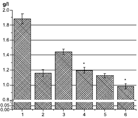
Актуальність. Підвищений рівень холестерину ліпопротеїнів високої щільності (ЛПВЩ) та аполіпопротеїну А1 (ApoA1) у плазмі асоціюється із зниженим ризиком розвитку серцево-судинних захворювань. Окрім потенційної кардіопротекторної функції, ЛПВЩ та ApoA1, основні аполіпопротеїни ЛПВЩ, також мають протидіабетичні властивості. Метою дослідження було визначити рівень ApoA1 у крові пацієнтів (n = 81) із цукровим діабетом (ЦД) та COVID-19. Матеріали та методи. Уміст ApoA1 визначали за допомогою наборів для імуноферментного аналізу (Elabscience, США). Вимірювання проводили при оптичній щільності 450 нм. Результати. Рівень ApoA1 у крові хворих на цукровий діабет і особливо на COVID-19 був значно нижчим, ніж у здорових людей. Дослідження залежності вмісту ApoA1 у плазмі від рівня Hb1Ac, статі та типу ЦД показало, що в крові хворих на ЦД 2-го типу кількість ApoA1 нижча, ніж в осіб із ЦД 1-го типу, і зі збільшенням рівня HbA1c уміст ApoA1 зменшується. Також була вірогідною гендерна різниця. Зі збільшенням індексу маси тіла рівень ApoA1 у плазмі крові зменшується нижче норми — 0,9 г/л, а при індексі маси тіла < 25 кг/м2 кількість ApoA1 значно перевищує середній рівень ліпопротеїну у хворих на ЦД. У пацієнтів із вперше діагностованим ЦД рівень ApoA1 значно вищий, а у пацієнтів із хворобою понад 10 років — нижчий від середнього та норми. Лікування бігуанідами в поєднанні з іншими препаратами (переважно інсуліном) або у вигляді монотерапії суттєво не впливає на вміст ApoA1 порівняно із середнім показником для всієї групи. У пацієнтів, які отримували похідні сульфонілсечовини, рівень ApoA1 значно нижчий від середнього для групи та від норми. Значний позитивний вплив на кількість ApoA1 у плазмі спостерігався в пацієнтів, які отримували комбінацію препаратів з інгібіторами натрійзалежного котранспортера глюкози 2-го типу, інсуліном та особливо інгібіторами дипептидилпептидази-4. Однак монотерапія інсуліном суттєво не впливала на вміст ApoA1. Обговорюються можливі механізми зниження ApoA1 за умов COVID-19 та ЦД. Висновки. Рівень ApoA1 може бути одним із перспективних маркерів тяжкого перебігу COVID-19.

Background. Increased level of high-density lipoprotein (HDL) cholesterol and apolipoprotein A1 (ApoA1) in plasma is associated with a reduced risk of developing cardiovascular diseases. In addition to its potential cardioprotective function, HDL and ApoA1, the main HDL apolipoprotein, also have antidiabetic properties. The aim of the study was to determine the level of ApoA1 in the blood of patients (n = 81) with diabetes mellitus and COVID-19. Materials and methods. ApoA1 was determined by enzyme-linked immunosorbent assay kits (Elabscience, USA). The measurements were performed at an optical density of 450 nm. Results. ApoA1 level in the blood of patients with diabetes and especially with COVID-19 was significantly lower than in healthy people. The study of the dependence of plasma ApoA1 content on the level of Hb1Ac, the gender and the type of diabetes showed that in blood of patients with type 2 diabetes the amount of ApoA1 is lower than in those with type 1 diabetes, and with an increase in the level of Hb1Ac the amount of ApoA1 decreases. There was also significant gender difference. With an increase in the body mass index, the content of ApoA1 in blood plasma decreases below normal — 0.9 g/L, and at body mass index < 25 kg/m2, the amount of ApoA1 is significantly higher than the average lipoprotein level in diabetic patients. In individuals with newly diagnosed diabetes, the level of ApoA1 is significantly higher, and in patients with more than 10 years of illness, it is below average and below normal. Biguanide treatment, either in combination with other drugs (mainly insulin) or as monothe-rapy, does not significantly affect the level of ApoA1 compared to the entire group average. In patients treated with sulfonylurea, the level of ApoA1 is significantly lower than the average level for the group and the norm. A significant positive effect on the amount of ApoA1 in plasma was observed in people treated with a combination of drugs with sodium-glucose cotransporter type 2 inhibitors, insulin and especially dipeptidyl peptidase-4 inhibitors. However, insulin monotherapy did not significantly affect the ApoA1 content. Possible mechanisms of ApoA1 decrease in COVID-19 and diabetes are discussed. Conclusions. Thus, the level of ApoA1 may be one of the promising markers of severe COVID-19.


Ключевые слова

аполіпопротеїн А1; COVID-19; цукровий діабет; серцево-судинні захворювання; гіпоглікемічні засоби

apolipoprotein A1; COVID-19; diabetes mellitus; cardiovascular diseases; hypoglycemic agents

Introduction

Apolipoprotein A1 (ApoA1), the main protein component of high-density lipoproteins (HDL), is a 243 amino acid polypeptide with an apparent moleculаr weight of 28 kDa. Circulating HDL particles contain single or multiple copies of ApoA1 [1]. ApoA1 is synthesized predominantly in the liver and small intestine [2]. Besides its role in HDL structure, ApoA1 is аlso critical for HDL functionality. ApoA1 in lipid-free form and in the nascent lipid-poor form — preβ1-HDL (consists mainly of ApoA1 and phospholipids with the molecular weight of 60–70 kDa) promotes efflux of cholesterol via the ATP-binding cassette transporter A1 (ABCA1) from macrophage foam cells and thus initiates the reverse cholesterol transport pathway from these cells, which is followed by facilitated hepatic uptake and ultimately excretion of the macrophage-derived cholesterol by the gut [1, 3]. Lipid-poor ApoA1 particles are abundant in interstitial fluids, where they can accept excess cholesterol from cholesterol-loaded cells. Recent data suggest that, by regulating cellular cholesterol homeostasis, HDL and ApoA1 can also regulate inflammatory responses in endothelial cells and other types of cells that have been activated by proinflammatory stimuli in the arterial intima [3, 4]. It has been found that increased levels of HDL-cholesterol (HDL-C) and ApoA1 in plasma are associated with a reduced risk of developing cardiovascular disease (CVD). In addition to its potential cardioprotective function, HDL and ApoA1 also have antidiabetic properties. Increases in plasma HDL and ApoA1 levels improve glycemic control in patients with type 2 diabetes mellitus by enhancing pancreatic β-cell function and improving insulin sensitivity, suggesting that interventions, which raise HDL levels, may be beneficial in diabetes-associated CVD [5, 6]. ApoA1 also stimulates glucose uptake in vivo into skeletal and cardiac muscles [7]. The ApoB/ApoA1 ratio has been found to be associated with type 2 diabetes and has been proposed as a novel biomarker for its prediction [8]. Meta-analysis also shows that decreased ApoA1 and increased ApoB levels, as well as the ApoB/A1 ratio, are risk factors for a first ischemic stroke [9].
Several clinical trials using HDL/ApoA1 infusion therapy have shown encouraging results. The use of gene transfer is an alternative way to exploit beneficial cardiovascular effects of HDL/ApoA1 in addition to HDL infusion therapy [10].

Materials and methods

The study was conducted at the Diabetology department of the Institute. The study protocol was approved by the Institute’s ethics committee (protocol 2, 15.02.2021). All patients signed informed consent to conduct further diagnostic and research study.
Blood was obtained by standard venipuncture and stored in EDTA vacutainer tubes. Plasma was separated by centrifugation within 10 min after blood sampling. The samples were stored at –80 °C until use. ApoA1 was determined (n = 81) using enzyme-linked immunosorbent assay kit (Elabscience, USA). The measurement was carried out at an optical density of 450 nm on the immunoenzymatic plate analyzer Stat Fax 3200 (Awareness Technology, USA).
Glycated hemoglobin was determined using one HbA1c FS kit (DiaSys Diagnostic Systems GmbH, Germany). The measurement was carried out at an optical density of 660 nm.
Statistical analysis and data presentation were performed using Origin 7.0 software. The results of the study are presented as M ± m. To compare the data groups, Student’s t-test was used. Values of P ≤ 0.05 were considered significant.

Results

The blood plasma of 60 type 2 diabetes patients and 21 patients (13 women and 8 men) with diabetes and COVID-19 was used. As a control, we used the blood of healthy people (n = 7) without concomitant diseases, of representative age. The level of HbA1c in diabetic patients was 9.62 ± 0.27 %; body mass index (BMI) — 30.69 ± 1.06 kg/m2.
As seen in Fig. 1, the average level of ApoA1 in the blood of healthy people is 1.88 g/l, which is close to the upper normal range (2.02–2.25 g/l). In diabetic patients, this indicator is significantly lower — 1.21 g/l, closer to the lower limit of the norm (1.04–1.08 g/l), and in diabetic patients after recovery from COVID-19, it is lower than the norm (Fig. 1, col. 2, 3). In patients with diabetes and COVID-19, the content of ApoA1 in the blood is approximately 0.25 g/l, which is more than 4 times lower than normal values. Interestingly, there are no differences between people with COVID-19 and diabetes, COVID-19 and CVD and patients with only COVID-19 (Fig. 1). In the blood of some individuals with COVID-19, the level of ApoA1 decreased to almost zero values — 0.09 g/l. The fact that the level of ApoA1 in the blood does not decrease in patients with COVID-19 and diabetes compared to those without diabetes indicates that COVID-19 creates significantly more powerful factors affecting the content of ApoA1, and such a decrease reaches a lower limit. It should be noted that after recovery from COVID-19, the level of ApoA1 restores, although it remains below the level in diabetic patients (Fig. 1, col. 2, 3).
One of the important indicators in diabetes is the level of HbA1c. The study of the dependence of plasma ApoA1 content on the level of HbA1c, the gender of patients and the type of diabetes showed that in type 2 diabetes the amount of ApoA1 is lower than in type 1, and with an increase in the level of HbA1c the amount of ApoA1 decreases. Gender differences were also significant (Fig. 2).
Body mass index is also an important indicator in diabetes. Fig. 3 shows that with an increase in BMI, the content of ApoA1 in blood plasma decreases below normal — 0.9 g/L, and at BMI < 25 kg/m2, the amount of ApoA1 is significantly higher than the average lipoprotein level in diabetic patients (Fig. 3, col. 2, 3).
The duration of the disease also affects the content of ApoA1 in the blood plasma (Fig. 4). In patients with newly diagnosed diabetes, the level of ApoA1 is significantly higher, and in those with more than 10 years of illness, it is below average and below normal (col. 2, 3, 6).
The association of diabetes duration and glycemic control (by HbA1c level) with the risks of CVD and all-cause mortality was firmly established. Both longer diabetes duration and poorer glycemic control were associated with elevated risks of CVD and mortality.
The ApoA1 level is influenced by the treatment method (Fig. 5). Biguanide treatment, either in combination with other drugs (mainly insulin) or as monotherapy, does not significantly affect the level of ApoA1 compared to the group average (col. 2, 3, 9). In patients treated with sulfonylurea, the level of ApoA1 is significantly lower than the average level for the entire group and the norm (col. 4). A significant positive effect on the amount of ApoA1 in plasma was observed in patients treated with a combination of drugs with sodium-glucose cotransporter type 2 inhibitors (iSGLT2), dipeptidyl peptidase-4 (DPP-4) inhibitors and insulin (col. 5–7). However, insulin monotherapy did not significantly affect the ApoA1 content (col. 8).

Discussion

In patients with insulin resistance and type 2 diabetes, plasma lipid and lipoprotein abnormalities are common [11]. A decrease in the ApoA1 blood level of diabetic patients and an increase in the CVD risk were noted in many studies [12–14]. The association of serum HDL-C and ApoA1 levels with risk of severe SARS-CoV-2 infection is established [15]. ApoA1 was identified as the protein complex seed, and amyloid beta A4 protein, epidermal growth factor, and complement C3 were the main bottlenecks in the network. ApoA1 carries anti-inflammatory properties that could assist in the regulation of the immune response. The obtained data indicate that upregulation of C3 and downregulation of ApoA1 in urine affect respiratory rigidity and thus the severity of COVID-19 [16]. Moreover, dysregulation of amyloid beta A4 protein and ApoA1 may contribute to the possible side effects of COVID-19 on the nervous system [17].
Lipid profiles were significantly affected by COVID-19 with decreased total cholesterol, HDL-C аnd low-density lipoprotein (LDL) cholesterol levels and increased triglyceride concentration compared to control subjects. Plasma ApoA1 was decreased by 55 % in patients versus controls [18]. COVID-19-induced hypolipidemia positively correlated with the severity of disease [19]. Decreased ApoA1 levels in COVID-19 patients suggest decreased synthesis by the liver and/or its replacement by serum amyloid A in HDL [18]. A decrease in ApoA1 was also associated with the pathogenesis of chronic hypersensitivity pneumonitis in terms of pulmonary fibrosis and mast cell chymase attenuated the protective effect of ApoA1 against pulmonary fibrosis. These results suggest that chymase produced by mast cells may play an important role in the degradation of ApoA1 [2, 20]. In addition, carboxypeptidase A and matrix metalloproteinases 3 and 14 can be involved in the degradation of ApoA1 [20, 21]. Inflammatory cytokines such as tumor necrosis factor and interleukin 1β, which are secreted in large quantities during COVID-19 infection, suppress the production of ApoA1 from hepatocytes and increase the expression of serum amyloid A, which becomes the major protein component of HDL in this context [10]. Finally, the regulation of ApoA1 expression can occur at the transcriptional level [22].
HbA1c was found to have significant positive correlation with total cholesterol, LDL-C, and triglyceride and significant negative correlation with HDL-C and HDL/LDL ratio [23]. Subjects with HbA1c-defined prediabetes and type 2 diabetes, respectively, are characterized by abnormalities in lipid profile — lower ApoA1 and HDL cholesterol levels [24].
It was shown that larger BMI, higher glucose levels, and lower content of ApoA1 are significantly and independently associated with newly diagnosed type 2 diabetes. Lower ApoA1 improved the risk prediction of new newly diagnosed 2 diabetes when it was added to the existing risk models [25]. Also, obesity, especially central obesity, contributes more to increasing ApoB/ApoA1 ratio than increased blood pressure, and other indices in women with polycystic ovary syndrome aged 20–38 years [26]. Obesity is associated with a state of chronic inflammation and increased cardiometabolic disease risk. All biomarkers were significantly associated with BMI: ApoA1, HDL-C, and 25(OH)D were inversely associated with BMI [27].
Clinicians should consider not only glycemic control but also diabetes duration in CVD risk assessments for participants with diabetes [28].
A significant decrease in the level of ApoA1 compared to its average content in diabetic patients after combined treatment with sulfonylurea is of particular interest. Some studies suggest that sulfonylureas may affect cardiac function and also may be associated with poorer outcomes after myocardial infarction [29]. Increased mortality from cardiovascular disease in diabetic patients taking tolbutamide was reported in the past decades. In the Mayo Clinic, in 185 consecutive diabetic patients undergoing percutaneous coronary intervention after myocardial infarction, the odds ratio for death was 2.77 for those treated with a sulfonylurea at the time of the myocardial infarction [29, 30]. Besides, high dose (500 μM) of glibenclamide inhibited ABCA1 function and ApoA1-mediated cholesterol efflux, and attenuated ABCA1 expression [31] that can lead to the accumulation of cholesterol in macrophages of atherosclerotic plaques. A decrease in the level of ApoA1 may be a reflection of the negative processes taking place during the treatment with sulfonylurea.
On the contrary, combination of drugs with iSGLT2 and DPP-4 inhibitors caused a small but significant increase in the level of ApoA1, which is consistent with their positive effect on cardiovascular function in diabetes [32, 33]. Moreover, glucagon-like peptide-1 receptor agonist, DPP-4 inhibitors and iSGLT2 can improve diabetic dyslipidemia [34].
Biguanide treatment, either in combination with other drugs (mainly insulin) or as monotherapy, does not significantly affect the level of ApoA1. The combined treatment with insulin and other drugs had a small positive effect on the level of ApoA1. It is known that hyperinsulinemia is an atherogenic factor [35], but treatment with insulin in combination with DPP-4 and iSGLT2 may lead to an increase in the plasma amount of ApoA1. The positive effect of metformin and especially the combination of metformin and insulin on the cardiovascular system may be explained by a decrease in endothelin-1 and NT-proBNP concentrations and by an increase in glucagon-like peptide-1 [36, 37].

Conclusions

ApoA1 level in the blood of patients with diabetes and especially with COVID-19 was significantly lower than in the blood of healthy people. The content of ApoA1 may be one of the promising markers of severe COVID-19.
The study of the dependence of the ApoA1 plasma content on the Hb1Ac level, the patients’ gender and the type of diabetes showed that in blood of individuals with type 2 diabetes, the ApoA1 amount is lower than in those with type 1 diabetes, and with an increase in the level of Hb1Ac the amount of ApoA1 decreases. There was also significant gender difference.
With an increase in BMI, the ApoA1 content in blood plasma decreases below normal — 0.9 g/l, and at BMI < 25 kg/m2, the amount of ApoA1 is significantly higher than the average lipoprotein level in diabetic patients.
In patients with newly diagnosed diabetes, the level of ApoA1 is significantly higher, and in people with more than 10 years of illness, it is below average and below normal.
Biguanide treatment, either in combination with other drugs (mainly insulin) or as monotherapy, does not significantly affect the level of ApoA1 compared to the entire group average. In patients treated with sulfonylurea, the level of ApoA1 is significantly lower than the average level for the group and the norm. A significant positive effect on the amount of ApoA1 in plasma was observed in patients treated with a combination of drugs with iSGLT2, insulin and especially DPP-4 inhibitors. However, insulin monotherapy did not significantly affect the ApoA1 content.
Possible mechanisms of ApoA1 decrease in COVID-19 and diabetes are discussed.
 
Received 21.06.2021
Revised 06.07.2021
Accepted 02.08.2021

Список литературы

  1. Lund-Katz S., Phillips M.C. High density lipoprotein structure-function and role in reverse cholesterol transport. Subcell. Biochem. 2010. 51. 183-227. doi: 10.1007/978-90-481-8622-8_7.
  2. Inoue Y., Okamoto T., Honda T. et al. Disruption in the ba-lance between apolipoprotein A-I and mast cell chymase in chronic hypersensitivity pneumonitis. Immun. Inflamm. Dis. 2020. 8(4). 659-671. doi: 10.1002/iid3.355.
  3. Kareinen I., Baumann M., Nguyen S.D. et al. Chymase released from hypoxia-activated cardiac mast cells cleaves human apoA-I at Tyr192 and compromises its cardioprotective activity. J. Lipid Res. 2018. 59(6). 945-957. doi: 10.1194/jlr.M077503.
  4. Mineo C., Shaul P.W. Regulation of signal transduction by HDL. J. Lipid Res. 2013. 54(9). 2315-24. doi: 10.1194/jlr.R039479.
  5. Rye K.A., Barter P.J., Cochran B.J. Apolipoprotein A-I interactions with insulin secretion and production. Curr. Opin. Lipidol. 2016. 27(1). 8-13. doi: 10.1097/MOL.0000000000000253.
  6. Di Bartolo B.A., Cartland S.P., Genner S. et al. HDL improves cholesterol and glucose homeostasis and reduces atherosclerosis in diabetes-associated atherosclerosis. J. Diabetes Res. 2021. 2021. 6668506. doi: 10.1155/2021/6668506.
  7. Fritzen A.M., Domingo-Espín J., Lundsgaard A.M. et al. ApoA-1 improves glucose tolerance by increasing glucose uptake into heart and skeletal muscle independently of AMPKα2. Mol. Metab. 2020. 35. 100949. doi: 10.1016/j.molmet.2020.01.013.
  8. Mao Y., Xu Y., Lu L. The nonlinear association between apolipoprotein B to apolipoprotein A1 ratio and type 2 diabetes. Medicine (Baltimore). 2017. 96(1). e5834. doi: 10.1097/MD.0000000000005834.
  9. Dong H., Chen W., Wang X. et al. Apolipoprotein A1, B le-vels, and their ratio and the risk of a first stroke: a meta-analysis and case-control study. Metab. Brain Dis. 2015. 30(6). 1319-1330. doi: 10.1007/s11011-015-9732-7.
  10. Chyu K.Y., Shah P.K. HDL/ApoA-1 infusion and ApoA-1 gene therapy in atherosclerosis. Front. Pharmacol. 2015. 6. 187. doi: 10.3389/fphar.2015.00187.
  11. Wolkowicz P., White C.R., Anantharamaiah G.M. Apolipoprotein mimetic peptides: an emerging therapy against diabetic inflammation and dyslipidemia. Biomolecules. 2021. 11(5). 627. doi: 10.3390/biom11050627.
  12. Cochran B.J., Ong K.L., Manandhar B., Rye K.A. High density lipoproteins and diabetes. Cells. 2021. 10(4). 850. doi: 10.3390/cells10040850.
  13. Gao L., Zhang Y., Wang X., Dong H. Association of apolipoproteins A1 and B with type 2 diabetes and fasting blood glucose: a cross-sectional study. BMC Endocr. Disord. 2021. 21(1). 59. doi: 10.1186/s12902-021-00726-5.
  14. Retnakaran R., Ye C., Connelly P.W., Hanley A.J., Sermer M., Zinman B. Serum apoA1 (apolipoprotein A-1), insulin resistance, and the risk of gestational diabetes mellitus in human pregnancy — brief report. Arterioscler. Thromb. Vasc. Biol. 2019. 39(10). 2192-2197. doi: 10.1161/ATVBAHA.119.313195.
  15. Hilser J.R., Han Y., Biswas S. et al. Association of serum HDL-cholesterol and apolipoprotein A1 levels with risk of severe SARS-CoV-2 infection. J. Lipid Res. 2021. 62. 100061. doi: 10.1016/j.jlr.2021.100061.
  16. Zamanian Azodi M., Arjmand B., Zali A., Razzaghi M. Introducing APOA1 as a key protein in COVID-19 infection: a bioinformatics approach. Gastroenterol. Hepatol. Bed Bench. 2020 Fall. 13(4). 367-373.
  17. Yang Y., Zhu Z., Fan L. et al. Low serum level of apolipoprotein A1 is an indicator of severity in patients with coronavirus disease 2019. Preprint. doi: 10.21203/rs.3.rs-31251/v1.
  18. Begue F., Tanaka S., Mouktadi Z. et al. Altered high-density lipoprotein composition and functions during severe COVID-19. Sci. Rep. 2021. 11(1). 2291. doi: 10.1038/s41598-021-81638-1.
  19. Wei X., Zeng W., Su J. et al. Hypolipidemia is associated with the severity of COVID-19. J. Clin. Lipidol. 2020. 14(3). 297-304. doi: 10.1016/j.jacl.2020.04.008.
  20. Usami Y., Kobayashi Y., Kameda T. et al. Identification of sites in apolipoprotein A-I susceptible to chymase and carboxypeptidase A digestion. Biosci. Rep. 2012. 33(1). 49-56. doi: 10.1042/BSR20120094.
  21. Park J.H., Park S.M., Park K.H., Cho K.H., Lee S.T. Analysis of apolipoprotein A-I as a substrate for matrix metalloproteinase-14. Biochem. Biophys. Res. Commun. 2011. 409(1). 58-63. doi: 10.1016/j.bbrc.2011.04.105.
  22. Georgila K., Vyrla D., Drakos E. Apolipoprotein A-I (ApoA-I), immunity, inflammation and cancer. Cancers (Basel). 2019. 11(8). 1097. doi: 10.3390/cancers11081097.
  23. Koval S.M., Yushko K.O., Snihurska I.O., Starchenko T.G., Pankiv V.I., Lytvynova O.M., Mysnychenko O.V. Relations of angiotensin-(1-7) with hemodynamic and cardiac structural and functional parameters in patients with hypertension and type 2 diabetes. Arterial Hypertension (Poland). 2019. 23(3). 183-189. doi: 10.5603/AH.a2019.0012.
  24. Calanna S., Scicali R., Di Pino A. et al. Lipid and liver abnormalities in haemoglobin A1c-defined prediabetes and type 2 diabetes. Nutr. Metab. Cardiovasc. Dis. 2014. 24(6). 670-676. doi: 10.1016/j.numecd.2014.01.013.
  25. Wu X., Yu Z., Su W. et al. Low levels of ApoA1 improve risk prediction of type 2 diabetes mellitus. J. Clin. Lipidol. 2017. 11(2). 362-368. doi: 10.1016/j.jacl.2017.01.009.
  26. Zheng J., Yin Q., Cao J., Zhang B. Obesity contributes more to increasing ApoB/ApoA1 ratio than hyperandrogenism in PCOS women aged 20–38 years in China. Exp. Ther. Med. 2017. 13(4). 1337-1342. doi: 10.3892/etm.2017.4094.
  27. Da Costa L.A., Arora P., García-Bailo B., Karmali M., El-Sohemy A., Badawi A. The association between obesity, cardiometabolic disease biomarkers, and innate immunity-related inflammation in Canadian adults. Diabetes Metab. Syndr. Obes. 2012. 5. 347-355. doi: 10.2147/DMSO.S35115.
  28. Li F.R., Yang H.L., Zhou R. et al. Diabetes duration and glycaemic control as predictors of cardiovascular disease and mortality. Diabetes Obes. Metab. 2021. 23(6). 1361-1370. doi: 10.1111/dom.14348.
  29. Sola D., Rossi L., Schianca G.P. et al. Sulfonylureas and their use in clinical practice. Arch. Med. Sci. 2015. 11(4). 840-848. doi: 10.5114/aoms.2015.53304.
  30. Garratt K.N., Brady P.A., Hassinger N.L., Grill D.E., Terzic A., Holmes D.R. Jr. Sulfonylurea drugs increase early mortality in patients with diabetes mellitus after direct angioplasty for acute myocardial infarction. J. Am. Coll. Cardiol. 1999. 33(1). 119-24. doi: 10.1016/s0735-1097(98)00557-9.
  31. Terao Y., Ayaori M., Ogura M. et al. Effect of sulfonylurea agents on reverse cholesterol transport in vitro and vivo. J. Atheroscler. Thromb. 2011. 18(6). 513-530. doi: 10.5551/jat.7641.
  32. Davies M.J., D’Alessio D.A., Fradkin J. et al. Management of hyperglycemia in type 2 diabetes, 2018. A consensus report by the American Diabetes Association (ADA) and the European Association for the Study of Diabetes (EASD). Diabetes Care. 2018. 41(12). 2669-2701. doi: 10.2337/dci18-0033.
  33. Scheen A.J. The safety of gliptins: updated data in 2018. Expert Opin. Drug Saf. 2018. 17(4). 387-405. doi: 10.1080/14740338.2018.1444027.
  34. Patti A.M., Giglio R.V., Papanas N., Rizzo M., Rizvi A.A. Future perspectives of the pharmacological management of diabetic dyslipidemia. Expert Rev. Clin. Pharmacol. 2019. 12(2). 129-143. doi: 10.1080/17512433.2019.1567328.
  35. Golshahi J., Validi E., Akbari M. The association between fasting serum insulin, apo-lipoproteins level, and severity of coronary artery involvement in non-diabetic patients. Adv. Biomed. Res. 2014. 3. 192. doi: 10.4103/2277-9175.140624.
  36. Sokolova L.K., Belchina Yu.B., Pushkarev V.V., Chervia-kova S.A., Vatseba T.S., Kovzun O.I., Pushkarev V.M., Tronko M.D. The effect of metformin treatment on the level of GLP-1, NT-proBNP and endothelin-1 in patients with type 2 diabetes mellitus. International Journal of Endocrinology (Ukraine). 2020. 16(8). 26-31. doi: 10.22141/2224-0721.16.8.2020.222882.
  37. Sokolova L.K., Belchina Y.B., Pushkarev V.V., Chervia-kova S.A., Vatseba T.S., Kovzun O.I., Pushkarev V.M. The level of endothelin-1 in the blood of patients with diabetes, treated with hypoglycemic drugs. Endokrynologia. 2020. 25(3). 201-206. doi: 10.31793/1680-1466.2020.25-3.201.

Вернуться к номеру