
Журнал "Гастроэнтерология" Том 57, №1, 2023
Вернуться к номеру
Характеристики стану підшлункової залози й печінки при хронічному панкреатиті на тлі пролікованого хронічного вірусного гепатиту С залежно від результатів тестування за міжнародною анкетою CAGE
Авторы: Бабінець Л.С., Хомин Г.О., Галабіцька І.М.
Тернопільський національний медичний університет імені І.Я. Горбачевського МОЗ України, м. Тернопіль, Україна
Рубрики: Гастроэнтерология
Разделы: Клинические исследования
Версия для печати
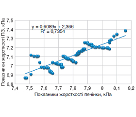
Актуальність. Встановлено, що в 35–56 % пацієнтів захворювання гепатобіліарної системи вважають фактором, що зумовлює формування або загострення хронічного панкреатиту. Також встановлено зменшення частки панкреатиту алкогольної етіології. Роль алкогольного фактора і його врахування при формуванні лікувальної тактики при ХП і коморбідності з хронічним вірусним гепатитом С (ХВГС) також потребує уточнення, адже відомо, що надмірне вживання алкоголю і куріння підвищує ризик розвитку панкреатиту у 8–17 разів. Мета дослідження: провести порівняльний аналіз параметрів структурного і функціонального стану печінки й підшлункової залози в пацієнтів із хронічним панкреатитом у коморбідності з пролікованим етіотропно хронічним вірусним гепатитом С залежно від результатів тестування за міжнародною анкетою CAGE. Матеріали та методи. Було обстежено 100 амбулаторних пацієнтів із ХП і супутнім ХВГС, пролікованим етіотропно. Усі хворі були обстежені за загальноприйнятими алгоритмами. Для встановлення впливу алкоголю на формування ХП і стан пацієнтів із пролікованим ХВГС верифікували прихований потяг до алкоголю, використовуючи міжнародну анкету CAGE. Вивчення щільності паренхіми печінки і ПЗ у пацієнтів проводили не тільки за даними ультразвукового дослідження у В-режимі, одночасно виконували вимірювання методом зсувнохвильової еластографії на скануючому ультразвуковому апараті Ultima PA з подальшим визначенням медіани параметрів у кілопаскалях (кПа). Визначення наявності й глибини зовнішньосекреторної недостатності підшлункової залози (ЗСНПЗ) проводили за вмістом фекальної еластази 1, який встановлювали імуноферментним методом. Результати. Скринінг-тестування пацієнтів із ХП на тлі пролікованого етіотропно ХВГС за шкалою CAGE дозволило констатувати, що прихований потяг до алкоголю мали 65,0 % таких хворих, причому 21,0 % цієї когорти становили жінки, що потребує урахування у веденні таких пацієнтів. Доведено, що в групі пацієнтів із CAGE ≥ 2,0 рівень функціональних і структурних змін печінки та ПЗ був вірогідно вищим (поглиблення ЗСНПЗ за зниженням фекальної α-еластази на 13,01 %, збільшення сумарного показника копрограми на 15,11 % і сумарного ультразвукового показника структури ПЗ — на 28,06 %, а сумарного ультразвукового показника структури печінки — на 40,68 % (р < 0,05)) і відповідав середньому ступеню тяжкості процесу в ПЗ за критеріями Марсельсько-Кембриджської класифікації, а у групі із CAGE < 2,0 — лише легкому ступеню. Висновки. Було доведено негативний вплив фактора посилення вживання алкоголю за CAGE за збільшенням щільності ехоструктури ПЗ на 5,73 % (р < 0,05), а печінки — на 5,16 % (р < 0,05). За результатами кореляційного аналізу залежності структурного стану печінки і ПЗ досліджуваних пацієнтів від значення шкали CAGE, які становили відповідно R = 0,713; р < 0,05, і R = 0,686; р < 0,05, констатували наявність сильних прямих залежностей структурного стану печінки і ПЗ від значення анкети CAGE, що довело самостійну вірогідно значиму роль вживання алкоголю для пацієнтів з коморбідним перебігом ХП і ХВГС.
Background. It was found that in 35–56 % of patients, hepatobiliary diseases are considered to be a factor causing the formation or exacerbation of chronic pancreatitis (CP) while reducing the share of pancreatitis of alcoholic etiology, as it was previously believed. However, the role of the alcohol factor and its consideration in the treatment strategy for CP and comorbidity with chronic viral hepatitis C (HCV) also needs to be clarified, because it is known that excessive alcohol consumption and smoking increases the risk of developing pancreatitis by 8–17 times. The purpose is to conduct a comparative analysis of parameters of the structural and functional state of the liver and pancreas in patients with chronic pancreatitis associated with chronic viral hepatitis C treated etiotropically, depending on the results of testing according to the international CAGE questionnaire. Materials and methods. We have examined 100 ambulatory patients with CP and concomitant HCV, treated etiotropically. All patients were examined according to generally accepted algorithms. To clarify the effect of alcohol on the formation of CP and the state of patients with treated HCV, latent craving for alcohol was verified using the international CAGE questionnaire. The study on the density of the liver parenchyma and the pancreas was carried out not only according to the ultrasound data in the B-mode, but also with the simultaneous shear wave elastography on the Ultima PA scanning ultrasound device with the further measurement of the median of the parameters in kilopascals. Determination of the presence and depth of exocrine pancreatic insufficiency was carried out by the content of fecal elastase-1 evaluated using the enzyme immunoassay method. Results. Screening-testing of patients with CP on the background of HCV treated etiotropically using the CAGE scale made it possible to state that 65.0 % of them had a latent craving for alcohol, and 21.0 % of this cohort were women, which needs to be taken into account in the management of such patients. It has been proven that in the group with CAGE ≥ 2.0, functional and structural changes in the liver and pancreas were significantly more severe (deepening of the exocrine pancreatic insufficiency with a decrease in fecal α-elastase by 13.01 %, an increase in the total index of the coprogram by 15.11 % and the total ultrasound indicator of the pancreatic structure by 28.06 %, and the total ultrasound indicator of the liver structure by 40.68 % (р < 0.05)) and corresponded to the average degree of severity of the process in the pancreas according to the criteria of the Marseille-Cambridge classification, and in the group with CAGE < 2.0 — only to a mild degree. Conclusions. The negative effect of the factor of increased alcohol use according to the CAGE was proven by increased density of pancreatic echotexture by 5.73 % (p < 0.05), and of the hepatic echotexture by 5.16 % (p < 0.05). According to the results of the correlation analysis on the dependence of the structural state of the liver and pancreas on the CAGE score, which were R = 0.713, p < 0.05, and R = 0.686, p < 0.05, respectively, there was found a strong direct dependence of the structural state of the liver and pancreas on the CAGE score, which proved an independent, reliably significant role of alcohol consumption for patients with a comorbid course of CP and HCV.
хронічний панкреатит; хронічний вірусний гепатит С; прихований потяг до алкоголю за анкетою CAGE; еластометрія хвилі зсуву; зовнішньосекреторна недостатність підшлункової залози
chronic pancreatitis; chronic viral hepatitis C; latent craving for alcohol according to the CAGE questionnaire; shear wave elastography; exocrine pancreatic insufficiency
Для ознакомления с полным содержанием статьи необходимо оформить подписку на журнал.
- Babinets L.S. et al. The influence of exocrine pancreatic insufficiency in the formation of osteopenia in patients with primary osteoarthritis. Wiadomosci lekarskie. 2020. P. 2238-2240.
- Babinets L.S., Sasyk H.M., Halabitska I.M., Mykuliak V.R. Possibilities of complex rehabilitation of patients with type 2 diabetes and concomitant chronic pancreatitis in ambulatory practice. Balneologia. 2021. Vol. 1. P. 12-15.
- Babinets L.S., Shevchenko N.A., Sasyk H.M., Zemlyak O.S. Research of lipid exchange diffusion of medium-age patients with chronic pancreatitis with diabetes mellitus. Journal of Education, Health and Sport formerly journal of Health Sciences. 2017. Vol. 7. 11. P. 309-318.
- Barr R.G., Ferraioli G., Palmeri M.L. Elastography assessment of liver fibrosis: society of radiologists in ultrasound consensus confe–rence statement. Radiology. 2015. Vol. 276. P. 845-861. doi: 10.1148/radiol.2015150619.
- Bitton S., Pettei M.J. Exocrine pancreatic insufficiency. Pediatr Rev. 2016. Vol. 37(2). P. 85-7. doi: 10.1542/pir.2015-0084.
- Capurso G., Traini M., Piciucchi M., Signoretti M. Exocrine pancreatic insufficiency: prevalence, diagnosis, and management. Arcidiacono. Clin Exp Gastroenterol. 2019. Vol. 21(12). P. 129-139. doi: 10.2147/CEG.S168266.
- Conwell D.L., Wu B.U. Chronic Pancreatitis: making the diag–nosis. Gastroenterol Hepatol. 2012. Vol. 10(10). P. 1088-95. doi: 10.1016/j.cgh.2012.05.015.
- Cote G.A. Alcohol and smoking as risk factors in an epidemio–logy study of patients with chronic pancreatitis. Clin Gastroenterol Hepatol. 2011. Vol. 9. P. 266-73. doi: 10.1016/j.cgh.2010.10.015.
- De la Iglesia D. et al. Pancreatic exocrine insufficiency and cardiovascular risk in patients with chronic pancreatitis: A prospective, longitudinal cohort study. J Gastroenterol Hepatol. 2019. Vol. 34(l). P. 277-283. doi: 10.1111/jgh. 14460.
- Dominguez-Munoz J.E. et al. Recommendations from the United European Gastroenterology evidence-based guidelines for the diagnosis and therapy of chronic pancreatitis. Pancreatology. 2018. Vol. 18(8). P. 847-54. doi: 10.1016/j.pan. 2018.09.016.
- Kothari D., Ketwaroo G., Freedman S.D., Sheth S.G. The Impact of risk factors of chronic pancreatitis on secretin pancreatic function testing: results of a 20-year study. Pancreas. 2017. Vol. 6(7). P. 887-90. doi:10.1097/MPA.0000000000000871.
- Lindkvist B. et al. Serum nutritional markers for prediction of pancreatic exocrine insufficiency in chronic pancreatitis. Pancreato–logy. 2012. Vol. 12. P. 305-10. doi: 10.1016/j.pan.2012.04.006.
- Lohr J.M., Oliver M.R., Frulloni L. Synopsis of recent guidelines on pancreatic exocrine insufficiency. United European Gastroenterol J. 2013. Vol. l(2). P. 79-83. doi: 10.1177/2050640613476500.
- Mitra S., De A., Chowdhury A. Epidemiology of non-alcoholic and alcoholic fatty liver diseases. Chowdhury. Transl Gastroenterol Hepatol. 2020. Vol. 5. P. 16. doi: 10.21037/tgh.2019.09.08.
- Pham A., Forsmark C. Chronic pancreatitis: review and update of etiology, risk factors, and management. FlOOORes. 2018. Vol.17. P. 7. doi: 10.12688/flresearch. 12852.1.
- Rodrigues-Pinto E. et al. Clube Portugues do Pancreas Re–commendations for Chronic Pancreatitis: Etiology, Natural History, and Diagnosis (Part 1). GE Port J Gastroenterol. 2019. Vol. 26(5). P. 346-55. doi: 10.1159/000497388.
- Samokhvalov A.V., Rehm J., Roerecke M. Alcohol consumption as a risk factor for acute and chronic pancreatitis: a systematic review and a series of meta-analyses. EBioMedicine. 2015 Nov 14. 2(12). 1996-2002. doi: 10.1016/j.ebiom.2015.11.023.
- Singhvi A., Yadav D. Myths and realities about alcohol and smoking in chronic pancreatitis. Curr Opin Gastroenterol. 2018. Vol. 34(5). P. 355-361. doi: 10.1097/MOG.0000000000000466.
- Westbrook R.H., Dusheiko G. Natural history of hepatitis C. J Hepatol. 2014. Vol. 61. S. 58-S68. doi: 10.1016/j.jhep.2014.07.012.
- Witt H., Apte M.V., Keim V., Wilson J.S. Chronic pancreatitis: challenges and advances in pathogenesis, genetics, diagnosis, and therapy. Gastroenterology. 2017. Vol. 132. P. 1557-73. doi: 10.1053/j.gastro.2007.03.001.
- Whitcomb D.C. Primer on precision medicine for complex chronic disorders. Clin Trans Gastroenterol. 2019. Vol. 10(7). e00067. doi: 10.14309/ctg.0000000000000067.
- Redkva O.V., Halabitska I.M. Evaluation of parameters of actual typical pathogenetic syndromes in comorbidity of type 2 diabetes mellitus and chronic pancreatitis. Wiad Lek. 2021. Vol. 74(10 cz 2). P. 2557-2559.
- Yang D., Forsmark C.E. Chronic pancreatitis. Curr Opin Gastroenterol. 2017. Vol. 33(5). P. 396-403. doi: 10.1097/MOG.0000000000000377.
- Ferraioli G. Review of liver elastography guidelines. J. Ultrasound Med. 2019. Vol. 38. P. 9-14. doi: 10.1002/jum.14856.
- Ferraioli G., Wong V.W., Castera L. Liver ultrasound elasto–graphy: an update to the world federation for ultrasound in medicine and biology guidelines and recommendations. Ultrasound Med Biol. 2018. Vol. 44. P. 2419-2440. doi: 10.1016/j.ultrasmedbio.2018.07.008.
- Foncea C.G. et al. The role of elastography for liver fibrosis screening in alcoholic liver disease. Med Ultrason. 2022. Vol. 24(4). P. 406-413. doi: 10.11152/mu-3784.
- Gianni E., Forte P., Galli V. Prospective evaluation of liver stiffness using transient elastography in alcoholic patients following abstinence. Alcohol. 2017. Vol. 52. P. 42-47. doi: 10.1093/alcalc/agw053.
- Mózes F.E., Lee J.A., Selvaraj E.A. Diagnostic accuracy of non-invasive tests for advanced fibrosis in patients with NAFLD: an individual patient data meta-analysis. Gut. 2022. Vol. 71. P. 1006-1019. doi: 10.1136/gutjnl-2021-324243.
- Barr R.G., Wilson S.R., Rubens D. Update to the society of radiologists in ultrasound liver elastography consensus statement. Radiology. 2020. Vol. 296. P. 263-274. doi: 10.1148/radiol.2020192437.
- Pose E., Ginès P. Transient elastography for alcoholic liver disease: a step forward. Lancet Gastroenterol Hepatol. 2018. Vol. 3. P. 589-591. doi: 10.1016/S2468-1253(18)30212-7.
- European Association for the Study of the Liver. EASL clinical practice guidelines on non-invasive tests for evaluation of liver di–sease severity and prognosis — 2021 update. J Hepatol. 2021. Vol. 75. P. 659-689. doi: 10.1016/j.jhep.2021.05.025.
- Singer M.V. Classification of pancreatitis — comparison of the revised 1984 Marseille Classification and the 1983 Cambridge classification. Comparative Study Z Gastroenterol Verh. 1991. Vol. 26. P. 39-42.
- Kahl M., Keller J., Layer P. Diagnosis: Functional testing, radiological work-up of chronic pancreatitis. Diseases of the Pancreas: Current Surgical Therapy. 2008. Р. 319-330.
- Sarner M., Cotton P.B. Classification of pancreatitis. Gut. 1984. Vol. 25(7). P. 756-759.
- Rumack C.M., Levine D. Diagnostic Ultrasound, 2-Volume Set. 5th Edition. Elsevier. 2017.