
Журнал «Медицина неотложных состояний» Том 19, №5, 2023
Вернуться к номеру
Ектопічні варикозно розширені вени: анатомічні особливості для хірургів і інтервенційних радіологів
Авторы: Чуклін С.М. (1), Чуклін С.С. (1), Чукла Р.О. (2)
(1) — Медичний центр Святої Параскеви, м. Львів, Україна
(2) — Львівський національний медичний університет імені Данила Галицького, м. Львів, Україна
Рубрики: Медицина неотложных состояний
Разделы: Справочник специалиста
Версия для печати
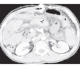
Ектопічний варикоз визначається як розширення портосистемних колатеральних вен у незвичних місцях, окрім гастроезофагеальної ділянки. Він розвивається внаслідок портальної гіпертензії, хірургічних втручань, аномалій венозного відтоку або тромбозу судин черевної порожнини і може мати сімейне походження. Ектопічні варикозні вени становлять клінічну проблему, оскільки їх важко локалізувати. Пропускання або неправильне тлумачення цих уражень може мати серйозні наслідки, а варіанти їх лікування неясні. Ектопічний варикоз можна виявити під час панендоскопії, ентероскопії, ендоскопічного ультразвукового дослідження, бездротової капсульної ендоскопії, діагностичної ангіографії, багатозрізової спіральної комп’ютерної томографії, магнітно-резонансної ангіографії, кольорової доплерографії, лапаротомії, лапароскопії, а іноді під час автопсії. Ектопічні варикозні вени можуть бути важливою причиною кровотечі та печінкової енцефалопатії, тому радіологи повинні ефективно їх ідентифікувати, щоб допомогти в прийнятті терапевтичного рішення. Знання анатомії та перебігу цих незвичайних портосистемних колатералей також має велике значення для інтервенційних радіологів і хірургів, бо дозволяє уникнути ненавмисного пошкодження судин під час інвазивних втручань. У цій статті ми розглядаємо частини шлунково-кишкового тракту й органи, які можуть бути залучені до ектопічного варикозу. Пошук літератури здійснювався в базі даних MedLine на платформі PubMed.
Ectopic varices are defined as dilated portosystemic collateral veins located in unusual sites, other than the gastroesophageal region. They develop secondary to portal hypertension, surgical procedures, anomalies in venous outflow, or abdominal vascular thrombosis and may be familial. Ectopic varices represent a clinical challenge because they are difficult to localize. Missing or misinterpreting these lesions can have serious consequences, and treatment options are unclear. Ectopic varices may be detected during panendoscopy, enteroscopy, endoscopic ultrasound, wireless capsule endoscopy, diagnostic angiography, multislice helical computed tomography, magnetic resonance angiography, color Doppler flow imaging, laparotomy, laparoscopy and occasionally during autopsy. They can be an important cause of bleeding and hepatic encephalopathy, so radiologists must effectively identify them to assist in making therapeutic decisions. Knowledge of the anatomy and course of these unusual portosystemic collaterals is also important for interventional radiologists and surgeons as it helps avoid inadvertent vascular damage during invasive procedures. In this article, we explore the parts of the gastrointestinal tract and organs that may be involved in ectopic varices. Literature search was conducted in the MedLine database on the PubMed platform.
ектопічні варикозно розширені вени; локалізація; анатомія; діагностика
ectopic varices; localization; anatomy; diagnosis
Для ознакомления с полным содержанием статьи необходимо оформить подписку на журнал.
- Simonetto D.A., Liu M., Kamath P.S. Portal Hypertension and Related Complications: Diagnosis and Management. Mayo Clin. Proc. 2019. 94(4). 714-726. doi: 10.1016/j.mayocp.2018.12.020.
- Sharma M., Rameshbabu C.S. Collateral pathways in portal hypertension. J. Clin. Exp. Hepatol. 2012. 2(4). 338-352. doi: 10.1016/j.jceh.2012.08.001.
- Henseler K.P., Pozniak M.A., Lee F.T. Jr, Winter T.C. 3rd. Three-dimensional CT angiography of spontaneous portosystemic shunts. Radiographics. 2001. 21(3). 691-704. doi: 10.1148/radiographics.21.3.g01ma14691.
- Ito K., Fujita T., Shimizu A., Sasaki K., Tanabe M., Matsunaga N. Imaging findings of unusual intra and extrahepatic portosystemic collaterals. Clin. Radiol. 2009. 64(2). 200-207. doi: 10.1016/j.crad.2008.05.016.
- Gebrael J., Yu H., Hyslop W.B. Spontaneous portoazygos shunt in a patient with portal hypertension. J. Radiol. Case Rep. 2013. 7(7). 32-36. doi: 10.3941/jrcr.v7i7.1437.
- Philips C.A., Anand L., Kumar K.N., Kasana V., Arora A. Rare, spontaneous trans-splenic shunt and intra-splenic collaterals with attendant splenic artery aneurysms in an adult patient with compensated cirrhosis and portal hypertension. Gastroenterol. Rep. (Oxf.). 2015. 3(2). 162-166. doi: 10.1093/gastro/gou047.
- Menon K.V., Kamath P.S. Regional and systemic hemodynamic disturbances in cirrhosis. Clin. Liver Dis. 2001. 5(3). 617-627. doi: 10.1016/s1089-3261(05)70184-7.
- Norton I.D., Andrews J.C., Kamath P.S. Management of ectopic varices. Hepatology. 1998. 28(4). 1154-1158. doi: 10.1002/hep.510280434.
- Moubarak E., Bouvier A., Boursier J. et al. Portosystemic collateral vessels in liver cirrhosis: a three-dimensional MDCT pictorial review. Abdom. Imaging. 2012. 37(5). 746-766. doi: 10.1007/s00261-011-9811-0.
- Hashimoto N., Akhoshi T., Yoshida D. et al. The efficacy of balloon-occluded retrograde transvenous obliteration on small intestinal variceal bleeding. Surgery. 2010. 148(1). 145-150. doi: 10.1016/j.surg.2009.10.052.
- Kocher N., Tripathy D., McAvoy N.C., Ireland H., Redhead D.N., Hayes P.C. Bleeding ectopic varices in cirrhosis: the role of transjugular intra-hepatic portosystemic stent shunts. Aliment. Pharmacol. Ther. 2008. 28(3). 294-303. doi: 10.1111/j.1365-2036.2008.03719.x.
- Onozato Y., Kakizaki S., Lizuka H. et al. Ectopic varices rupture in the gastroduodenal anastomosis successfully treated with Nbutyl-2-cyanoacrylate injection. Acta Med. Okayama. 2007. 61(6). 361-365. doi: 10.18926/AMO/32880.
- Hashizumi M., Tanoue K., Ohta M. et al. Vascular anatomy of duodenal varices: angiographic and histopathological assessment. Am. J. Gastroenterol. 1993. 88(11). 1942-1945. PMID: 8237946.
- Saad W.E., Lippert A., Saad N.E., Caldwell S. Ectopic varices: anatomical classification, hemodynamic classification, and hemodynamic-based management. Tech. Vasc. Interv. Radiol. 2013. 16(2). 158-175. doi: 10.1053/j.tvir.2013.02.004.
- Sato T. Transabdominal color Doppler ultrasonography for the diagnosis of small intestinal and vesical varices in a patient successfully treated with percutaneous transhepatic obliteration. Clin. J. Gastroenterol. 2010. 3(4). 214-218. doi: 10.1007/s12328-010-0164-y.
- Hashizume M., Kitano S., Sugimachi K., Sueishi K. Three-dimensional view of the vascular structure of the lower esophagus in clinical portal hypertension. Hepatology. 1988. 8(6). 1482-1487. doi: 10.1002/hep.1840080603.
- Amin R., Alexix R., Kowzis J. Fatal ruptured duodenal varix: a case report and review of literature. Am. J. Gastroenterol. 1985. 80(1). 13-18. PMID: 3871304.
- Khouqeer F., Morrow C., Jordan P. Duodenal varices as a cause of massive upper gastrointestinal bleeding. Surgery. 1987. 102(3). 548-552. PMID: 3498234.
- Lebrec D., Behamou J. Ectopic varices in portal hypertension. Clin. Gastroenterol. 1985. 14(1). 105-121. PMID: 3872747.
- Sharma M., Rameshbabu C.S., Garg S., Rai P. Portal venous system and its tributaries: a radial endosonographic assessment. Endosc. Ultrasound. 2012. 1(2). 96-107. doi: 10.7178/eus.02.008.
- Arora A., Rajesh S., Meenakshi Y.S., Sureka B., Bansal K., Sarin S.K. Spectrum of hepatofugal collateral pathways in portal hypertension: an illustrated radiological review. Insights Imaging. 2015. 6(5). 559-572. doi: 10.1007/s13244-015-0419-8.
- McAlister V.C., Al-Saleh N.A. Duodenal dearterialization and stapling for severe hemorrhage from duodenal varices with portal vein thrombosis. Am. J. Surg. 2005. 189(1). 49-52. doi: 10.1016/j.amjsurg.2004.04.011.
- Yi F., Guo X., Zeng Q.L. et al. Computed Tomography Images of Spontaneous Portosystemic Shunt in Liver Cirrhosis. Can. J. Gastroenterol. Hepatol. 2022. 2022. 3231144. doi: 10.1155/2022/3231144.
- Ibukuro K., Tsukiyama T., Mori K., Inoue Y. Veins of Retzius at CT during arterial portography: anatomy and clinical importance. Radiology. 1998. 209(3). 793-800. doi: 10.1148/radiology.209.3.9844676.
- Lim L.G., Lee Y.M., Tan L., Chang S., Lim S.G. Percutaneous paraumbilical embolization as an unconventional and successful treatment for bleeding jejunal varices. World J. Gastroenterol. 2009. 15(30). 3823-3826. doi: 10.3748/wjg.15.3823.
- Francois F., Tadros C., Diehl D. Pan-colonic varices and idiopathic portal hypertension. J. Gastrointestin. Liver Dis. 2007. 16(3). 325-328. PMID: 17925930.
- Weingart J., Höchter W., Ottenjann R. Varices of the entire colon: an unusual cause of recurrent intestinal bleeding. Endoscopy. 1982. 14(2). 69-70. doi: 10.1055/s-2007-1021582.
- Ito K., Shiraki K., Sakai T., Yoshimura H., Nakano T. Portal hypertensive colopathy in patients with liver cirrhosis. World J. Gastroenterol. 2005. 11(20). 3127-3130. doi: 10.3748/wjg.v11.i20.3127.
- Philips C.A., Arora A., Shetty R., Kasana V. A Comprehensive Review of Portosystemic Collaterals in Cirrhosis: Historical Aspects, Anatomy, and Classifications. Int. J. Hepatol. 2016. 2016. 6170243. doi: 10.1155/2016/6170243.
- Dhiman R.K., Saraswat V.A., Choudhuri G., Sharma B.C., Pandey R., Naik S.R. Endosonographic, endoscopic and histologic evaluation of alterations in the rectal venous system in patients with portal hypertension. Gastrointest. Endosc. 1999. 49(2). 218-227. doi: 10.1016/s0016-5107(99)70490-6.
- Chawla Y., Dilawari J.B., Katariya S. Gallbladder wall varices in portal vein thrombosis. Am. J. Roentgenol. 1994. 162(3). 643-645. doi: 10.2214/ajr.162.3.8109513.
- Chandra R., Kapoor D., Tharakan A., Chaudhary A., Sarin S.K. Portal biliopathy. J. Gastroenterol. Hepatol. 2001. 16(10). 1086-1092. doi: 10.1046/j.1440-1746.2001.02562.x.
- Vellar I.D. Preliminary study of the anatomy of the venous drainage of the intrahepatic and extrahepatic bile ducts and its relevance to the practice of hepatobiliary surgery. ANZ J. Surg. 2001. 71(7). 418-422. doi: 10.1046/j.1440-1622.2001.02150.x.
- Sharma M., Pathak A. Perforators of common bile duct wall in portal hypertensive biliopathy (with videos). Gastrointest. Endosc. 2009. 70(5). 1041-1043. doi: 10.1016/j.gie.2009.03.040.
- Sharma M., Pathak A. Intracholedochal varices in portal hypertensive biliopathy. Eur. J. Radiol. Extra. 2009. 72(3). e119-e123. doi: 10.1016/j.ejrex.2009.06.001.
- Martin B.F., Tudor R.G. The umbilical and paraumbilical veins of man. J. Anat. 1980. 130(Pt 2). 305-322. PMID:7400038.
- Gaspar Y., Detry O., de Leval J. Vesical varices in a patient with portal hypertension. N. Engl. J. Med. 2001. 345(20). 1503-1504. doi: 10.1056/NEJM200111153452018.
- Loganathan G., Shyamkumar N.K., Kurian G. et al. Vesical varix in cirrhosis of liver. Indian J. Gastroenterol. 2000. 19(4). 193-194. PMID:11059194.
- McHugh P.P., Jeon H., Gedaly R., Johnston T.D., Depriest P.D., Ranjan D. Vaginal varices with massive hemorrhage in a patient with nonalcoholic steatohepatitis and portal hypertension: successful treatment with liver transplantation. Liver Transpl. 2008. 14(10). 1538-1540. doi: 10.1002/lt.21506.
- Helmy A., Al Kahtani K., Al Fadda M. Updates in the pathogenesis, diagnosis and management of ectopic varices. Hepatol. Int. 2008. 2(3). 322-334. doi: 10.1007/s12072-008-9074-1.
- Hashimoto M., Heianna J., Yasuda K. et al. Portal flow into the liver through veins at the site of biliary-enteric anastomosis. Eur. Radiol. 2005. 15(7). 1421-1425. doi: 10.1007/s00330-005-2667-3.
- Seeger M., Günther R., Hinrichsen H. et al. Chronic portal vein thrombosis: transcapsular hepatic collateral vessels and communicating ectopic varices. Radiology. 2010. 257(2). 568-578. doi: 10.1148/radiol.10100157.
- Mitchell A.W., Jackson J.E. Trans-anastomotic porto-portal varices in patients with gastrointestinal haemorrhage. Clin. Radiol. 2000. 55(3). 207-211. doi: 10.1053/crad.1999.0347. PMID: 10708614.
- Choi J.W., Lee C.H., Kim K.A., Park C.M., Kim J.Y. Ectopic varices in colonic stoma: MDCT findings. Korean J. Radiol. 2006. 7(4). 297-299. doi: 10.3348/kjr.2006.7.4.297.
- Araki T., Imaizumi A., Okada H., Sasaki Y., Onishi H. Anatomy of Left Inferior Phrenic Vein in Patients Without Portal Hypertension. Am. J. Roentgenol. 2021. 217(2). 411-417. doi: 10.2214/AJR.20.23106.
- Nagahiro I., Toda D., Andou A., Shimizu N. A case of bronchial varices due to extrahepatic portal hypertension. Respiration. 2007. 74(4). 460-461. doi: 10.1159/000088356.
- Agarwal A., Jain M. Multidetector CT portal veno-graphy in evaluation of portosystemic collateral vessels. J. Med. Imaging Radiat. Oncol. 2008. 52(1). 4-9. doi: 10.1111/j.1440-1673.2007.01903.x.
- Ikeda O., Nakasone Y., Yokoyama K., Inoue S., Takamori H., Baba H., Yamashita Y. Successful treatment of mesenteric varices by retrograde transvenous obliteration by the delivery of N-butyl-2-cyanoacrylate via an abdominal wall vein. Cardiovasc. Intervent. Radiol. 2014. 37(2). 533-536. doi: 10.1007/s00270-013-0647-6.
- Kang H.K., Jeong Y.Y., Choi J.H. et al. Three-dimensional multidetector row CT portal venography in the evaluation of portosystemic collateral vessels in liver cirrhosis. Radiogra-
- phics. 2002. 22(5). 1053-1061. doi: 10.1148/radiographics.22.5.g02se011053.
- Bataille L., Baillieux J., Remy P., Gustin R.M., Denié C. Spontaneous rupture of omental varices: an uncommon cause of hypovolemic shock in cirrhosis. Acta Gastroenterol. Belg. 2004. 67(4). 351-354. PMID: 15727080.