
Журнал «Медицина неотложных состояний» Том 19, №6, 2023
Вернуться к номеру
Перспективи вивчення деяких електрофізіологічних параметрів у діагностиці затримки росту плода
Авторы: Лахно І.В. (1), Мартиненко О.В. (2), Раймонді Ж. (3), Шульгін В.І. (4)
(1) — Харківський національний медичний університет, м. Харків, Україна
(2) — Харківський національний університет ім. В.Н. Каразіна, м. Харків, Україна
(3) — Університет Ла Сапієнца, м. Рим, Італія
(4) — Національний аерокосмічний університет ім. М.Є. Жуковського «Харківський авіаційний інститут»,
м. Харків, Україна
Рубрики: Медицина неотложных состояний
Разделы: Клинические исследования
Версия для печати
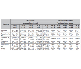
Актуальність. Затримка росту плода є клінічним синдромом, що має несприятливі проєкції як на перинатальні наслідки, так і на здоров’я людини в дорослому житті. Традиційні підходи до діагностики цього синдрому базуються на ультразвуковій біометрії та доплерометрії матково-плацентарного і плодово-пуповинного кровотоку. Апаратура, яка використовується для таких досліджень, є дуже дорогою, а підготовка кваліфікованого спеціаліста займає тривалий час. Неінвазивна електрокардіографія дає можливість оцінювати показники варіабельності серцевого ритму плода і морфологічні параметри комплексу pQRST. Метою роботи було встановлення показників варіабельності серцевого ритму і морфологічних параметрів за даними неінвазивної електрокардіографії плода, які можна використати для діагностики затримки росту плода. Матеріали та методи. У роботі взяли участь 220 вагітних жінок віком від 18 до 40 років у терміни 24–41 тиждень. Усіх обстежених пацієнток було розподілено на дві групи. До І групи було віднесено 108 жінок із затримкою росту плода, а до ІІ групи — 112 вагітних зі здоровими плодами. Реєстрацію електрокардіограми плода здійснювали в абдомінальному відведенні протягом 30–60 хвилин. Результати. Проведена робота дозволила встановити наявність статистично вірогідних змін деяких показників варіабельності серцевого ритму на тлі затримки росту плода: SI, TP, AC/DC, LF, співвідношення LF/HF, а також параметрів морфології: тривалості QTc, ширини комплексу QRS і співвідношення T/QRS. Отримані дані демонстрували порушення вегетативної регуляції на тлі затримки росту плода і відображали напругу регуляторних систем, а саме ступінь домінування активності центральних механізмів регуляції над автономними. Порушення процесів де- і реполяризації були наслідком ремоделювання міокарда плода під впливом ішемії. Висновки. Розробка алгоритмів діагностики затримки росту плода за допомогою неінвазивної електрокардіографії потребує подальшого тестування таких показників: SI, TP, AC/DC, LF, LF/HF, тривалості QTc, ширини комплексу QRS і співвідношення T/QRS.
Background. Fetal growth restriction is a clinical syndrome that has adverse effects on both perinatal outcomes and human health in adulthood. Traditional approaches to the diagnosis of this syndrome are based on ultrasound biometry and dopplerometry of uteroplacental and fetal-umbilical blood flow. The equipment used for such research is very expensive, and the training of a qualified specialist takes a long time. Non-invasive electrocardiography makes it possible to evaluate fetal heart rate variability and morphological parameters of the pQRST complex. The purpose: to determine indices of heart rate variability and morphological parameters based on the data of fetal non-invasive electrocardiography, which can be used to diagnose fetal growth restriction. Materials and methods. Two hundred and twenty pregnant women aged 18 to 40 years on 24–41 weeks of gestation were enrolled in the study. All of them were divided into two groups: 108 women with fetal growth restriction were included in group I, and 112 patients with healthy fetuses in group II. The fetal electrocardiogram was recorded in abdominal lead for 30–60 minutes. Results. The performed work made it possible to detect statistically significant changes in some indices of heart rate variability on the background of fetal growth restriction: SI, TP, AC/DC, LF, LF/HF ratio, as well as morphological parameters: QTc duration, QRS complex width and T/QRS ratio. The obtained data demonstrated a violation of autonomic regulation following fetal growth restriction and reflected the tension of regulatory systems, namely, the degree of dominance of the activity of central regulatory mechanisms over autonomous ones. Impaired de- and repolarization processes were a consequence of fetal myocardium remodeling due to ischemia. Conclusions. The development of algorithms for the diagnosis of fetal growth restriction using non-invasive electrocardiography requires further research of the following parameters: SI, TP, AC/DC, LF, LF/HF ratio, QTc duration, QRS complex width, and T/QRS ratio.
затримка росту плода; неінвазивна електрокардіографія плода; варіабельність серцевого ритму
fetal growth restriction; non-invasive fetal electrocar-diography; heart rate variability
Для ознакомления с полным содержанием статьи необходимо оформить подписку на журнал.
- Benítez Marín M.J., Blasco Alonso M., González Mesa E. Prenatal Predictors of Neurobehavioral Outcome in Children with Fetal Growth Restriction at 6 Years of Age: A Retrospective Cohort Study. Children (Basel). 2023 Jun 1. 10(6). 997. doi: 10.3390/children10060997.
- Kingdom J.C., Audette M.C., Hobson S.R., Windrim R.C., Morgen E. A placenta clinic approach to the diagnosis and management of fetal growth restriction. Am. J. Obstet. Gynecol. 2018 Feb. 218(2S). S803-S817. doi: 10.1016/j.ajog.2017.11.575.
- Nardozza L.M., Caetano A.C., Zamarian A.C. et al. Fetal growth restriction: current knowledge. Arch. Gynecol. Obstet. 2017 May. 295(5). 1061-1077. doi: 10.1007/s00404-017-4341-9.
- Lakhno I. The Insight into Obstetric Care near the Front Line in Kharkiv. Acta Med. Litu. 2022. 29(2). 236-44. doi: 10.15388/Amed.2022.29.2.10.
- Hoyer D., Żebrowski J., Cysarz D. et al. Monitoring fetal maturation-objectives, techniques and indices of autonomic function. Physiol. Meas. 2017 May. 38(5). R61-R88. doi: 10.1088/1361-6579/aa5fca.
- Stampalija T., Casati D., Monasta L., Sassi R., Rivolta M.W., Muggiasca M.L., Bauer A., Ferrazzi E. Brain sparing effect in growth-restricted fetuses is associated with decreased cardiac acceleration and deceleration capacities: a case-control study. BJOG. 2016 Nov. 123(12). 1947-1954. doi: 10.1111/1471-0528.13607.
- Hoyer D., Schmidt A., Gustafson K.M. et al. Heart rate variability categories of fluctuation amplitude and complexity: diagnostic markers of fetal development and its disturbances. Physiol. Meas. 2019 Jul 3. 40(6). 064002. doi: 10.1088/1361-6579/ab205f.
- Liu B., Ridder A., Smith V. et al. Feasibility of antenatal ambulatory fetal electrocardiography: a systematic review. J. Matern. Fetal. Neonatal. Med. 2023 Dec. 36(1). 2204390. doi: 10.1080/14767058.2023.2204390.
- Velayo C.L., Funamoto K., Silao J.N.I. et al. Evaluation of Abdominal Fetal Electrocardiography in Early Intrauterine Growth Restriction. Front. Physiol. 2017 Jun 26. 8. 437. doi: 10.3389/fphys.2017.00437.
- Fuchs T. Values of T/QRS ratio in pregnancies complicated by intrauterine growth restriction. J. Perinat. Med. 2016 Oct 1. 44(7). 751-757. doi: 10.1515/jpm-2014-0305.
- Lakhno I.V. The hemodynamic repercussions of the autonomic modulations in growth-restricted fetuses. Alexandria Journal of Medicine. 2017. 53 (4). 333-336. doi: 10.1016/j.ajme.2016.12.007.
- Zizzo A.R., Kirkegaard I., Uldbjerg N. et al. Towards better reliability in fetal heart rate variability using time domain and spectral domain analyses. A new method for assessing fetal neurological state? PLoS One. 2022 Mar 1. 17(3). e0263272. doi: 10.1371/journal.pone.0263272.
- Cao S., Xiao H., Gong G., Fang W., Chen C. Morphology extraction of fetal ECG using temporal CNN-based nonlinear adaptive noise cancelling. PLoS One. 2022 Dec 15. 17(12). e0278917. doi: 10.1371/journal.pone.0278917.
- Kahankova R., Martinek R., Jaros R., Behbehani K., Matonia A., Jezewski M., Behar J.A. A Review of Signal Processing Techniques for Non-Invasive Fetal Electrocardiography. IEEE Rev. Biomed. Eng. 2020. 13. 51-73. doi: 10.1109/RBME.2019.2938061.
- Nayak S.K., Bit A., Dey A. et al. A Review on the Nonlinear Dynamical System Analysis of Electrocardiogram Signal. J. Healthc. Eng. 2018 May 2. 2018. 6920420. doi: 10.1155/2018/6920420.
- Smith V., Nair A., Warty R., Sursas J.A., da Silva Costa F., Wallace E.M. A systematic review on the utility of non-invasive electrophysiological assessment in evaluating for intra uterine growth restriction. BMC Pregnancy Childbirth. 2019 Jul 5. 19(1). 230. doi: 10.1186/s12884-019-2357-9.
- Zizzo A.R., Hansen J., Peteren O.B. et al. Growth-restricted human fetuses have preserved respiratory sinus arrhythmia but reduced heart rate variability estimates of vagal activity during quiescence. Physiol. Rep. 2022 Nov. 10(22). e15458. doi: 10.14814/phy2.15458.
- Odendaal H.J., Kieser E., Crockart I.C. et al. Clinical associations of fetal heart rate accelerations as derived from transabdominal fetal electrocardiograms. Eur. J. Obstet. Gynecol. Reprod. Biol. 2022 Dec. 279. 5-11. doi: 10.1016/j.ejogrb.2022.10.005.
- Matonia A., Jezewski J., Kupka T., Jezewski M., Horoba K., Wrobel J., Czabanski R., Kahankowa R. Fetal electrocardiograms, direct and abdominal with reference heartbeat annotations. Sci. Data. 2020 Jun 25. 7(1). 200. doi: 10.1038/s41597-020-0538-z.
- Aisa M.C., Cappuccini B., Favilli A. et al. Biochemical and Anthropometric Parameters for the Early Recognition of the Intrauterine Growth Restriction and Preterm Neonates at Risk of Impaired Neurodevelopment. Int. J. Mol. Sci. 2023 Jul 17. 24(14). 11549. doi: 10.3390/ijms241411549.
- Stroux L., Redman C.W., Georgieva A. et al. Doppler-based fetal heart rate analysis markers for the detection of early intrauterine growth restriction. Acta. Obstet. Gynecol. Scand. 2017 Nov. 96(11). 1322-1329. doi: 10.1111/aogs.13228.
- Kapaya H., Jacques R., Anumba D. Comparison of diurnal variations, gestational age and gender related differences in fetal heart rate (FHR) parameters between appropriate-for-gestational-age (AGA) and small-for-gestational-age (SGA) fetuses in the home environment. PLoS One. 2018 Mar 9. 13(3). e0193908. doi: 10.1371/journal.pone.0193908.
- Priante E., Verlato G., Giordano G. et al. Intrauterine Growth Restriction: New Insight from the Metabolomic Approach. Metabolites. 2019 Nov 6. 9(11). 267. doi: 10.3390/metabo9110267.
- Frasch M.G. Heart rate variability code: does it exist and can we hack it? Bioengineering (Basel). 2023 Jul 10. 10(7). 822. doi: 10.3390/bioengineering10070822.
- Zhang Y., Gu A., Xiao Z., Xing Y., Yang C., Li J., Liu C. Wearable Fetal ECG Monitoring System from Abdominal Electrocardiography Recording. Biosensors (Basel). 2022 Jun 30. 12(7). 475. doi: 10.3390/bios12070475.