
Журнал «Здоровье ребенка» Том 18, №7, 2023
Вернуться к номеру
Аналіз стану мікроциркуляторного русла у дітей з гострими бронхітами
Авторы: Буряк О.Г., Нечитайло Ю.М.
Буковинський державний медичний університет, м. Чернівці, Україн
Рубрики: Педиатрия/Неонатология
Разделы: Клинические исследования
Версия для печати
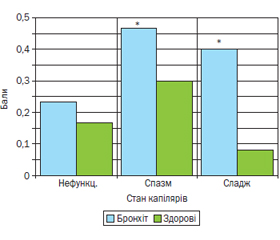
Мета дослідження. Оцінити особливості стану мікроциркуляторного русла (МЦР) за даними біомікроскопії при захворюваннях дихальної системи у дітей на моделі гострого бронхіту. Матеріали та методи. У роботі подано дані вивчення МЦР у дітей з гострим бронхітом (30 пацієнтів) порівняно зі здоровими особами. Стан мікроциркуляції реєстрували за допомогою біомікроскопії нігтьового ложа та оцінювали за якісними та напівкількісними показниками. Результати. У дітей з гострим бронхітом порівняно зі здоровими дітьми відмічено низку змін: в артеріолах — збільшення звивистості, у венулярних відділах — явища сладжу та спазмованості, у капілярах — зменшення кількості функціонуючих мікросудин та зміну їх форми, уповільнення кровотоку та сладжування еритроцитів. Висновки. Цифрова біомікроскопія судин нігтьового ложа у дітей є доступним, простим та інформативним методом вивчення стану МЦР при гострих респіраторних захворюваннях. При гострих бронхітах, навіть з нетяжким перебігом, спостерігаються зміни картини МЦР на рівні всіх мікросудин, з переважанням змін у капілярах.
Background. The purpose was to evaluate the peculiarities of microcirculation (MC) according to biomicroscopy data in respiratory diseases in children on a model of acute bronchitis. Materials and methods. The paper presents data on the study of MC in children with acute bronchitis (n = 30) compared with healthy individuals. The state of microcirculation was evaluated using nail bed biomicroscopy and by qualitative and semi-quantitative indicators. Results. In children with acute bronchitis compared to healthy persons, a number of changes were noted: in the arterioles — an increase in tortuosity, in the venular parts — the phenomena of sludge and spasms, in the capillaries — a decrease in the number of functioning microvessels and changes in their shape, slowing blood flow and sludging of red blood cells. Conclusions. Digital nail bed biomicroscopy in children is an accessible, simple and informative method for studying the state of MC in acute respiratory diseases. In acute bronchitis, even with a mild course, there are changes in the MC pattern at the level of all microvessels, with a predominance of changes in the capillaries.
діти; біомікроскопія; мікроциркуляція; гострий бронхіт
children; biomicroscopy; microcirculation; acute bronchitis
Для ознакомления с полным содержанием статьи необходимо оформить подписку на журнал.
- Bottari G., Damiani E., Confalone V., Scorcella C., Casarotta E., Gandolfo C., et al. Microvascular dysfunction in pediatric patients with SARS-COV-2 pneumonia: report of three severe cases. Microvasc. Res. 2022 May. 141. 104312. doi: 10.1016/j.mvr.2022.104312.
- Colantuoni A., Martini R., Caprari P., Ballestri M., Capecchi P.L., Gnasso A., et al. COVID-19 sepsis and microcirculation dysfunction. Front. Physiol. 2020 Jun 26. 11. 747. doi: 10.3389/fphys.2020.00747.
- Natalello G., De Luca G., Gigante L., et al. Nailfold capillaro–scopy findings in patients with coronavirus disease 2019: broadening the spectrum of COVID-19 microvascular involvement. Microvasc. Res. 2021 Jan. 133. 104071. doi: 10.1016/j.mvr.2020.104071.
- Low D.A., Jones H., Cable N. et al. Historical reviews of the assessment of human cardiovascular function: interrogation and understan–ding of the control of skin blood flow. Eur. J. Appl. Physiol. 2020. 120(1). 1-16. doi: 10.1007/s00421-019-04246-y.
- Corliss B.A., Mathews C., Doty R. et al. Methods to label, image, and analyze the complex structural architectures of microvascular networks. Microcirculation. 2019 Jul. 26(5). e12520. doi: 10.1111/micc.12520.
- McClatchey P.M., Mignemi N.A., Xu Z. et al. Automated quantification of microvascular perfusion. Microcirculation. 2018 Aug. 25(6). e12482. doi: 10.1111/micc.12482.
- Nechytailo Yu.M., Nechytailo D.Iu., Buriak O.H. Features of the condition of microcirculation in schoolage children with arterial hypertension. Austrian Journal of Technical and Natural Sciences. 2014. 9–10. 86-89.
- Bruel A., Bacchetta J., Ginhoux T., Rodier-Bonifas C., Sellier-Leclerc A.L., Fromy B., et al. Skin microvascular dysfunction as an early cardiovascular marker in primary hyperoxaluria type I. Pediatr. Nephrol. 2019 Feb. 34(2). 319-327. doi: 10.1007/s00467-018-4081-5.
- Ugenti V., Romano A.C., Tibirica E. Microvascular endothelial dysfunction during cardiopulmonary bypass in surgery for correction of cyanotic and acyanotic congenital heart disease. Microvasc. Res. 2018 Nov. 120. 55-58. doi: 10.1016/j.mvr.2018.06.004.
- Ocampo-Garza S.S., Villarreal-Alarcón M.A., Villarreal-Tre–viño A.V., Ocampo-Candiani J. Capillaroscopy: A Valuable Diagnostic Tool. Actas Dermosifiliogr. (Engl. Ed). 2019 Jun. 110(5). 347-352. English, Spanish. doi: 10.1016/j.ad.2018.10.018.
- Wright I.M., Latter J.L., Dyson R.M., Levi C.R., Clifton V.L. Videomicroscopy as a tool for investigation of the microcirculation in the newborn. Physiol. Rep. 2016 Oct. 4(19). e12941. doi: 10.14814/phy2.12941.
- Erdem Ö., Ince C., Tibboel D., Kuiper J.W. Assessing the Microcirculation with Handheld Vital Microscopy in Critically Ill Neonates and Children: Evolution of the Technique and Its Potential for Critical Care. Front. Pediatr. 2019 Jul 9. 7. 273. doi: 10.3389/fped.2019.00273.
- Tamez-Rivera O., Villarreal-Treviño A.V., Castañeda-Macazaga T., Britton-Robles S.C., Ramos-Gómez L.I., Rubio-Pérez N.E. Abnormal Nailfold Capillaroscopy in a Patient with Multisystem Inflammatory Syndrome in Children. Pediatr. Infect. Dis J. 2021 Mar 1. 40(3). e113-e115. doi: 10.1097/INF.0000000000003022.
- Deegan A.J., Wang R.K. Microvascular imaging of the skin. Phys. Med. Biol. 2019 Mar 21. 64(7). 07TR01. doi: 10.1088/1361-6560/ab03f1.
- Çakmak F., Demirbuga A., Demirkol D., Gümüş S., Torun S.H., Kayaalp G.K., et al. Nailfold capillaroscopy: A sensitive method for evaluating microvascular involvement in children with SARS-CoV-2 infection. Microvasc. Res. 2021 Nov. 138. 104196. doi: 10.1016/j.mvr.2021.104196.
- Low D.A., Jones H., Cable N. et al. Historical reviews of the assessment of human cardiovascular function: interrogation and understan–ding of the control of skin blood flow. Eur. J. Appl. Physiol. 2020. 120(1). 1-16. doi: 10.1007/s00421-019-04246-y.
- Corliss B.A., Mathews C., Doty R. et al. Methods to label, –image, and analyze the complex structural architectures of microvascular networks. Microcirculation. 2019 Jul. 26(5). e12520. doi: 10.1111/micc.12520.
- Hamilton N.B., Attwell D., Hall C.N. Pericyte-mediated regulation of capillary diameter: a component of neurovascular coupling in health and disease. Front Neuroenergetics. 2010; 2. 5. doi: 10.3389/fnene.2010.00005.
- Meijer E.M., van Dijk C.G.M., Kramann R., Verhaar M.C., Cheng C. Implementation of pericytes in vascular regeneration stra–tegies. Tissue Eng. Part B Rev. 2022. 28(1). 1-21. doi: 10.1089/ten.TEB.2020.0229.
- Ewerlöf M., Salerud E.G., Strömberg T., Larsson M. Estimation of skin microcirculatory hemoglobin oxygen saturation and red blood cell tissue fraction using a multispectral snapshot imaging system: a validation study. J. Biomed. Opt. 2021 Feb. 26(2). 026002. doi: 10.1117/1.JBO.26.2.026002.
- Bai Y., Li G., Yung L., Yu P.B., Ai X. Intrapulmonary arterial contraction assay reveals region-specific deregulation of vasoreactivity to lung injuries. Am. J. Physiol. Lung Cell Mol. Physiol. 2023 Aug 1. 325(2). L114-L124. doi: 10.1152/ajplung.00293.2022.
- Rayner S.G., Hung C.F., Liles W.C., Altemeier W.A. Lung pericytes as mediators of inflammation. Am. J. Physiol. Lung Cell Mol. Physiol. 2023 Jul 1. 325(1). L1-L8. doi: 10.1152/ajplung.00354.2022.