
Журнал «Здоровье ребенка» Том 18, №7, 2023
Вернуться к номеру
Клініко-морфологічна характеристика та діагностика зовнішніх гемангіом у дітей
Авторы: S. Toktosunova, A. Toktosunov, U. Sharshenov
I.K. Akhunbaev Kyrgyz State Medical Academy, Bishkek, Kyrgyz Republic
Рубрики: Педиатрия/Неонатология
Разделы: Клинические исследования
Версия для печати
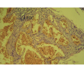
Актуальність. Актуальність теми полягає в тому, що в дітей віком до 1 року все частіше зустрічаються гемангіоми, коли необхідне підтвердження діагнозу морфологічними методами. Мета дослідження: охарактеризувати початкові та кінцеві гістологічні зміни гемангіом шкіри та представити основні клінічні прояви гемангіом у дітей. Матеріали та методи. У роботі використано декілька клінічних (для детальної діагностики захворювання), морфологічних (гістологічне дослідження матеріалу) та статистичних методів. Об’єктом дослідження були 98 дітей з гемангіомами шкіри обличчя віком від 6 місяців до 16 років. Результати. Проведене морфологічне дослідження показало, що в 49 випадках (50 %) виявлено типову просту капілярну структуру гемангіоми, у 35 випадках (35,7 %) — кавернозний тип новоутворення. Лише 18,3 % досліджуваних дітей мали комбіновані гемангіоми. Гемангіоми капілярного типу містять багато дрібних капілярів із вузьким просвітом, покритих спаяним епітелієм. Висновки. Під час фази активного росту гемангіоми спостерігається переважання масивних ендотеліальних клітин. Це свідчить про те, що основна та найбільша частина гіперплазії представлена судинним компонентом, зокрема проліферуючими капілярами, вистеленими епітелієм.
Background. The relevance lies in the increasing occurrence of haemangiomas in children under 1 year old when diagnosis confirmation through morphological methods is necessary. The purpose of the research is to characterise the histological initial and final changes of cutaneous haemangiomas and to present the main clinical manifestations of haemangiomas in children. Materials and methods. Several clinical (for detailed diagnosis of the disease), morphological (histological examination of the material) and statistical methods were used in the work. The object of the research was 98 children with haemangiomas of the facial skin aged 6 months to 16 years. Results. The morphological study demonstrated that in 49 cases (50 %), the typical simple capillary structure of haemangioma was identified, in 35 cases (35.7 %) — cavernous type of neoplasm. Only 18.3 % of the children had combined haemangiomas. Capillary-type haemangiomas contain many small capillaries with a narrow lumen, covered by adhering fleshy epithelium. Conclusions. During the phase of the active growth of haemangiomas, there is a predominance of massive endothelial cells, suggesting that the vascular component, particularly proliferating epithelium-lined capillaries, makes up the main and largest part of the hyperplasia.
шкіра; новоутворення; судинний компонент; вроджена патологія; кріодеструкція
skin; neoplasm; vascular component; congenital pathology; cryodestruction
Для ознакомления с полным содержанием статьи необходимо оформить подписку на журнал.
- Leung A.K.C., Lam J.M., Leong K.F., Hon K.L. Infantile he–mangioma: an updated review. Curr. Pediatr. Rev. 2021. 17(1). 55-69. doi: 10.2174/1573396316666200508100038.
- Rodríguez Bandera A.I., Sebaratnam D.F., Wargon O., Wong L.F. Infantile hemangioma. Part 1: Epidemiology, pathogenesis, clinical presentation and assessment. J. Am. Acad. Dermatol. 2021. 85(6). 1379-1392. doi: 10.1016/j.jaad.2021.08.019.
- Sebaratnam D.F., Rodríguez Bandera A.L., Wong L.F., Wargon O. Infantile hemangioma. Part 2: Management. J. Am. Acad. Dermatol. 2021. 85(6). 1395-1404. doi: 10.1016/j.jaad.2021.08.020.
- Mrad M.A., AlMarshad F.A., AlZahrani A.M., Obeid D.A., Alsinan T.A., AlOtaibi A.S. A cosmetic approach to cheek and nasal he–mangioma utilizing open rhinoplasty in a 10-year-old child: a case report. Cureus. 2023. 15(1). e34185. doi: 10.7759/cureus.34185.
- Tang L.N., Liu G., Yang C., Sha X.W., Wang S.Y. Short-term efficacy of superficial X-ray treatment for infantile maxillofacial he–mangioma. West China J. Stomatol. 2021. 39(4). 464-468. doi: 10.7518/hxkq.2021.04.014.
- Li C.X., Li H., Zhou Y.C., Gong Z.C., Ling B. Application of topical betaxolol to cure superficial infantile hemangioma: a pilot study. Pediatr. Int. 2022. 64(1). e15384. doi: 10.1111/ped.15384.
- Kim H.W., Yoo S.Y., Oh S., Jeon T.Y., Kim J.H. Ultrasonography of pediatric superficial soft tissue tumors and tumor-like lesions. Korean J. Radiol. 2020. 21(3). 341-355. doi: 10.3348/kjr.2019.0343.
- Berenguer B., Lorca-García C., Lancharro A., DeTomás E. Pediatric tumors of the buccal fat pad: lipoma and hemangioma. Acta Chir. Belg. 2020. 120(5). 341-343. doi: 10.1080/00015458.2019.1570743.
- Jiang J.C., Xu Q., Fang S., Gao Y., Jin W.W. Sequelae after involution of superficial infantile hemangioma: early intervention with –595-nm pulsed laser combined with 755-nm long-pulsed alexandrite laser versus wait-and-see. Clin. Cosmet. Invest. Dermatol. 2021. 14. 37-43. doi: 10.2147/CCID.S279140.
- He H.Y., Shi W.K., Jiang J.C., Gao Y., Xue X.M. An exploration of optimal time and safety of 595-nm pulsed dye laser for the treatment of early superficial infantile hemangioma. Dermatol. Ther. 2022. 35(5). e15406. doi: 10.1111/dth.15406.
- Dahan E., Abou Jaoude L. Infantile hemangiomas: a review of current treatment options. Pediatr. Ann. 2023. 52(5). 192-197. doi: 10.3928/19382359-20230307-02.
- Fan P.H., Liang D., Jia L.J., Gong Y.B., Sun B. et al. Clinicopathological features of verrucous hemangioma. Chinese J. Pathol. 2021. 50(12). 1341-1345. doi: 10.3760/cma.j.cn112151-20210602-00402.
- Bancalari B., Colmenero I., Noguera-Morel L., Torrelo A., Enríquez de Salamanca J., Hernández-Martín Á. Papillary hemangioma in a child and sonographic characterization. Pediatr. Dermatol. 2020. 37(1). 233-234. doi: 10.1111/pde.14048.
- Zheng H., Cai Y., Xu Y., Yu Q., Kang B. et al. Predictors of treatment responses of superficial infantile hemangiomas to topical timolol. Dermatol. Ther. 2022. 35(11). e15833. doi: 10.1111/dth.15833.
- Mazzini C., Vicini G., Nicolosi C., Pieretti G., Bacherini D. еt al. Multimodal imaging of a retinal cavernous hemangioma. Eur. J. Ophthalmol. 2022. 32(2). NP53-NP56. doi: 10.1177/1120672120971549.
- Kadriyan H., Sulaksana M.A., Yudhanto D., Aryani I.G.A.T., Yuliani E.A. et al. Subcutaneous hemangioma on nasal dorsum: a case report. J. Med. Case Rep. 2020. 14(1). 128. doi: 10.1186/s13256-020-02443-4.
- Liu Y.X., Zhang J., Nie X.L., Chen Y.L., Wang Z.Y., Xu Z.G. The effect of topical sirolimus on superficial Kaposiform haemangioendothelioma. Australas. J. Dermatol. 2021. 62(2). 329-331. doi: 10.1111/bjd.16400.
- Rikhotso R.E., Alharbi A.A. Management of refractory mandibular kaposiform hemangioendothelioma with sirolimus: a case report and review of the literature. J. Oral Maxillofac. Surg. 2021. 79(10). 2086-2088. doi: 10.1016/j.joms.2021.05.017.
- Ghirardo S., Cozzi G., Trevisan G., Addesa S., Berti I., Barbi E. Deep sedation for laser treatment of cutaneous vascular anomalies in children. Lasers Med. Sci. 2020. 35(7). 1649-1652. doi: 10.1007/s10103-020-02998-0.
- Lin Z., Zhang B., Yu Z., Li H. The effectiveness and safety of topical β-receptor blocker in treating superficial infantile haemangiomas: a meta-analysis including 20 studies. Br. J. Clin. Pharmacol. 2020. 86(2). 199-209. doi: 10.1111/bcp.14196.
- Nagata E., Kashiwagura Y., Okada E., Tanaka S., Sano S. et al. Efficacy and safety of propranolol cream in infantile hemangioma: a prospective pilot study. J. Pharmacol. Sci. 2022. 149(2). 60-65. doi: 10.1016/j.jphs.2022.03.004.
- Nakazono M., Kagimoto S., Koike T., Satake T., Maegawa J. Clinical outcomes of small infantile hemangiomas treated with pulsed dye laser. Dermatol. Surg. 2022. 48(8). 833-837. doi: 10.1097/DSS.0000000000003491.
- Gomes R., Salazar L., Fraga C., Correia M.R., Barbosa-Sequeira J. et al. Management of infantile hemangiomas — experience of a tertiary hospital. Eur. J. Pediatr. 2023. 182(4). 1611-1618. doi: 10.1007/s00431-023-04827-2.
- Geisler E.L., Brannen A., Pressler M., Perez J., Kane A.A., Hallac R.R. 3D imaging of vascular anomalies using raster-scanning optoacoustic mesoscopy. Lasers Surg. Med. 2022. 54(10). 1269-1277. doi: 10.1002/lsm.23588.
- Bouchghoul H., Benachi A., Senat M.V. Prenatal percutaneous fetoscopic laser photocoagulation of chorioangioma: report of two cases and review of the literature. Fetal Diagn. Ther. 2021. 48(8). 633-639. doi: 10.1159/000517392.
- Karkoska K., Ricci K., Vanden Heuvel K., Trout A.T., Smith E.A. et al. Metastatic neuroblastoma masquerading as infantile hemangioma in a 4-month-old child. Pediatr. Blood Cancer. 2021. 68(5). e28920. doi: 10.1002/pbc.28920.
- Tarmizi N.N.A., Bastion M.L.C., Aziz R.A.A., Din N.M., Khialdin S.M. Retinal racemose hemangioma with optic neuropathy in a child. Cureus. 2021. 13(8). e17620. doi: 10.7759/cureus.17620.
- Lin E.Y., Rao L., Wang X.H. Detection of superficial lymphatic malformation with dermoscopy and reflectance confocal microscopy. Skin Res. Technol. 2023. 29(2). e13283. doi: 10.1111/srt.13283.
- Lin Z. Reply to “Comment on “The effectiveness and safety of topical beta-receptor blocker in treating superficial infantile haemangiomas: a meta-analysis including 20 studies” Dr. Sun et al.”. Br. J. Clin. Pharmacol. 2022. 88(6). 3002-3003. doi: 10.1111/bcp.15246.
- Shi W., He H., Jiang J., Gao Y., Quan H. Timing and efficacy of 595-nm pulsed-dye laser combined with 0.5% timolol maleate solution in the treatment of superficial infantile hemangiomas. Clin. Cosmet. Invest. Dermatology. 2021. 14. 1593-1599. doi: 10.2147/CCID.S337850.
- Xie M., Liu J., Zhou P., Xu X., Liu H. et al. Development of a digital imaging analysis system to evaluate the treatment response in superficial infantile hemangiomas. PLoS ONE. 2023. 18(3). e0282274. doi: 10.1371/journal.pone.0282274.
- Chang S.J., Wang H., Chen J., Chen Q., Chang L. et al. Evaluation of transdermal transport and concurrent cutaneous hydrolysis of timolol prodrug for the treatment of infantile hemangiomas. Pharm. 2022. 14(8). 1607. doi: 10.3390/pharmaceutics14081607.
- Belmesk L., Dubois J., Caouette-Laberge L., David M., Bortoluzzi P. et al. Kaposiform hemangioendothelioma with overlapping features of rapidly involuting congenital hemangioma and a delayed complication of necrotizing fasciitis. Pediatr. Dermatol. 2020. 37(5). 902-906. doi: 10.1111/pde.14262.
- Tognetti L., Pianigiani E., Ierardi F., Cartocci A., Fiorani D. et al. A new clinical and dermoscopic monitoring of infantile hemangiomas treated with oral propranolol. Dermatol. Ther. 2020. 33(6). e14283. doi: 10.1111/dth.14283.
- Mashiah J., Bar-Ilan E., Koren A., Friedman O., Zur E., Artzi O. Enhanced percutaneous delivery of beta-blockers using thermal resurfacing drug delivery system for topical treatment of infantile hemangiomas. Dermatol. 2020. 236(6). 565-570. doi: 10.1159/000507808.
- Cheng J.W.C.H., Lam Y.Y., Fung G.P.G., Sin C., Luk D.C.K. et al. Randomised controlled trial: can topical timolol maleate prevent complications for small superficial infantile haemangiomata in high-risk areas? Pediatr. Res. 2020. 88(5). 756-760. doi: 10.1038/s41390-020-0917-3.
- Wang X., Feng W., Zhao X., Liu Z., Dong L. The efficacy and safety of topical β-blockers in treating infantile hemangiomas: a meta-analysis including 11 randomized controlled trials. Dermatology. 2021. 237(3). 433-443. doi: 10.1159/000510029.
- Ying H., Han Y., Chen Z., Yu Z., Chang L. et al. The effect of 0.5% topical timolol cream on regression of superficial infantile haemangioma in the involuting phase: a prospective, randomized, self-controlled study. Eur. J. Dermatol. 2020. 30(5). 591-595. doi: 10.1684/ejd.2020.3708.
- Cohen P.R. Tender cutaneous neoplasms: case reports of patients with a symptomatic dermatofibroma and a new acrostic for painful tumors of the skin. Cureus. 2022. 14(9). e29713. doi: 10.7759/cureus.29713.
- Diociaiuti A., Paolantonio G., Zama M., Alaggio R., Carnevale C. et al. Vascular birthmarks as a clue for complex and syndromic vascular anomalies. Front. Pediatr. 2021. 9. 7303. doi: 10.3389/fped.2021.730393.
- Jicha K.I., Wu S., Drake A.F., Morrell D.S. Hemangiomatous neck plaques in a child with cleft lip. Pediatr. Dermatol. 2021. 38(4). 929-931. doi: 10.1111/pde.14541.
- Rezende K.M., de Barros Gallo C., Nogueira G.P., Corraza A.C., Haddad A.E. et al. Retrospective study of oral lesions biopsied in babies and toddlers. Oral Dis. 2023. doi: 10.1111/odi.14552.
- Schapovalova O., Gorlova A., de Munter J., Sheveleva E., Eropkin M. et al. Immunomodulatory effects of new phytotherapy on human macrophages and TLR4- and TLR7/8-mediated viral-like inflammation in mice. Front. Med. 2022. 9. 952977. doi: 10.3389/fmed.2022.952977.
- Rubins A.Y., Branta D.K., Hartmane I.V., Rajevska A.S., Gutmane R.A., Lielbriedis Y.M. Multiple carcinoma in patients with psoriasis caused by psoralen-ultraviolet A therapy, natural killer cell insufficiency, or intensified sun exposure? Cutis. 1992. 49(6). 430-432.
- Sundaramoorthy D., Chandrasekaran A. Patient Dose Analysis Using GafchromicTM EBT3 Film: A Retrospective Study with a Four Dual-Field Technique in Total Skin Electron Therapy. Asian Pacific J. Cancer Prevent. 2023. 24(7). 2505-2513.
- Hartmane I., Mikažans I., Ivdra I., Derveniece A., Ančupane I. Experience of phototherapy in dermatological praxis in complex therapy of psoriasis patients. Proceed Latvian Acad. Sci. Sec. B: Natur. Exact Appl. Sci. 2016. 70(1). 7-12.
- Hajiyeva N., Gafarov I., Hajiyeva A., Sultanova N., Panahova T. Forecasting of atopic dermatitis in newborns. Indian J. Dermatol. 2022. 67(3). 311. doi: 10.4103/ijd.ijd_933_21.