
Журнал «Здоровье ребенка» Том 19, №1, 2024
Вернуться к номеру
Вірусні пневмонії у дітей дошкільного віку: актуальні аспекти клініко-параклінічної діагностики та лікування
Авторы: Мокія-Сербіна С.О., Литвинова Т.В., Шелевицька В.А., Заболотня Н.І.
Дніпровський державний медичний університет, м. Дніпро, Україна
Рубрики: Педиатрия/Неонатология
Разделы: Справочник специалиста
Версия для печати
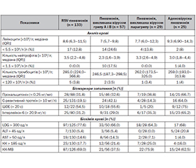
Актуальність. Стаття присвячена актуальним проблемам інфектології і педіатрії — вірусним пневмоніям. Вірусні інфекції можуть бути причиною численних порушень організму, проте ускладнення з боку дихальних шляхів є найбільш поширеними, особливо у дітей дошкільного віку. Мета: на основі узагальнення наявної літератури подати клініко-параклінічні особливості та лікування пневмоній, викликаних поширеними вірусними збудниками у дітей дошкільного віку. Матеріали та методи. Розглянуто і проаналізовано сучасні вітчизняні та зарубіжні літературні джерела щодо клініко-параклінічної діагностики та лікування пневмоній, викликаних поширеними вірусними збудниками. Для пошуку літературних джерел вивчались бази даних Web of Science, Scopus, PubMed, Google Scholar. При визначенні рекомендацій взято до уваги нову версію Міжнародної класифікації хвороб — 11 та результати проспективних і ретроспективних досліджень, проведених останніми роками. Результати. Наведено сучасні епдеміологічні та етіологічні дані щодо вірусних пневмоній у дітей, актуалізована їх термінологічна база, що сприяє закріпленню цього діагнозу в педіатричній практиці. У контексті проблеми, на основі аналізу результатів багатоцентрового ретроспективного обстеження, наведені відмінності в клінічних проявах, лабораторних показниках та результатах візуалізації при пневмоніях, викликаних поширеними вірусними збудниками. Висвітлено клінічні предиктори наявності коінфекцій мікоплазми та вторинної бактеріальної інфекції. Рекомендовано більш широке впровадження сучасних методів етіологічної діагностики через потенціал високої частки супутніх інфекцій у дітей з вірусною пневмонією. Розглянуто питання лікування пневмоній у дітей дошкільного віку. Висновки. Результати проведеного аналізу обґрунтовують наявність суттєвої залежності між видами збудника і особливостями перебігу вірусної пневмонії у дітей дошкільного віку, підвищують ефективність диференціальної діагностики різних клінічних фенотипів та оцінку лабораторної активності захворювання. Ці дані, з високим ступенем вірогідності, допоможуть забезпечити вибір оптимальної стартової терапії, прогнозувати перебіг захворювання і вирішити питання щодо необхідності госпіталізації.
Background. The article deals with the relevant problems of infectology pediatrics — viral pneumonias. Viral infections can be the cause of numerous disorders of the body, but respiratory complications are the most common, especially in preschool children. The purpose is to present the clinical and paraclinical features and treatment of pneumonia caused by common viral pathogens in preschool children based on a generalization of the available literature. Materials and methods. Modern domestic and foreign literary sources on clinical and paraclinical diagnosis and treatment of pneumonia caused by common viral pathogens were reviewed and analyzed. Web of Science, Scopus, PubMed, Google Scholar databases were studied to search for literature sources. When determining the recommendations, a new, 11th revision of the International Classification of Diseases and the results of prospective and retrospective studies conducted in recent years were taken into account. Results. Modern epidemiological and etiological data on viral pneumonias in children are presented, their terminology base is updated, which contributes to the consolidation of this diagnosis in pediatric practice. In the context of the problem, based on the analysis of the results of a multicenter retrospective examination, differences in clinical manifestations, laboratory parameters, and imaging results in pneumonia caused by common viral pathogens are presented. Clinical predictors for the presence of mycoplasma co-infections and secondary bacterial infection are highlighted. Wider implementation of modern methods of etiological diagnosis is recommended because of the risk of a high rate of co-infections in children with viral pneumonia. The issue of pneumonia treatment in preschool children is considered. Conclusions. The results of the analysis justify the existence of a significant dependence between the types of the pathogen and the features of viral pneumonia course in preschool children, increase the effectiveness of differential diagnosis in various clinical phenotypes and the assessment of laboratory activity of the disease. These data, with a high degree of probability, can ensure the choice of the optimal initial therapy, predict the disease course, and solve the issue of the need for hospitalization.
огляд; пневмонія; аденовірусна; респіраторно-синцитіальна; грипозна; парагрипозна; діагностика; діти
review; pneumonia; adenovirus, respiratory syncytial, influenza, parainfluenza diagnosis; children
Для ознакомления с полным содержанием статьи необходимо оформить подписку на журнал.
- Freeman A.M., Leigh T.R., Jr. Viral Pneumonia. In: StatPearls [Internet]. Treasure Island (FL): StatPearls Publishing; 2024 Jan. 2023 Jul 4. https://www.ncbi.nIm.nih.gov/books/NBK513286.
- Shoar S., Musher D.M. Etiology of Community-acquired pneumonia in adults: a systematic review. Pneumonia (Nathan). 2020. 12. 11. https//doi.org/10.1186/s41479-020-00074-3.
- Murdoch D.R., O’Brien K.L., Driscoll A.J., Karron R.A. Laboratory Methods for Determining Pneumonia Etiology in Children. Clinical Infectious Diseases. 2012. 54. Iss. suppl_2. S146-S152. https://doi.org/10.1093/cid/cir1073. https://academic.oup.com/cid/article/54/suppl 2/S146/376724.
- Palacios G., Hanig M., Cistan D., Savji N., Bussetti A.V. Streptococcus pneumonia coinfection is correlated with the severity of H1N1 pandemic influenza. Plos One. 2009. 4(12). e8540. https://doi.org/10.1371/jornal pone.0008540.
- Torres A., Lee N., Cilloniz C., Vila L., Van der Ferden M. Laboratory diagnosis of pneumonia in the molecular age. European Resperatori Journal. 2016. 48. 1764-1778. doi: 10.1183/1399300301144-2016. https://erj.ersjournals/com/content/48/6/1764.
- Revised WHO classification and treatment of pneumonia in children at health facilities: Evidence summaries. (2014). Geneva: World Health Organization (www.who.int/about/licensing/copyright_form/en/index.html).
- Sethi B.S. Aspiration Pneumonitis and pneumonia. URL: https:www.msd manuals. com/professional/pulmonary-disorders/pneumonia/aspiration-pneumonitis s-and pneumonia.
- Mauro C., Reynolds C.F., Maercher A. Prolonged grief disorder: clinical utility of ICG-11 diagnostik duidelenes. Psychol. Med. 2019. 49(5). 861-867. doi: 10.1017/s0033291718001563.
- Torres A., Cilloniz C., Niederman M.S. Pneumonia. Not. Rev. Dis. Primers. 2021. 7(1). 25. doi: 10.1038/s41572-021-00259-0.
- Dichson R.P., Erb-Downward J.R., Martinez F.J. The Microbiome and the Respiratory Tract. ARI. 2015 Oktober 22. 13. 48. doi: 10.1146/annure-physiol-021115-105238.
- Nathan A.M., Ju Teh C.S., Jabar K.A., Teong B.T., Thangaperumal A. Bacterial pneumonia and its associated factors in children from a developing country: A Prospective cohort study. doi: 10.1371/journall.pone.0228056; https://journall.plos.org/plosone/article.
- Ruedа Z.V., Aguilar Y., Maya M.A., Zopez L., Restepo A., Garces C. Etiology and the challenge of diagnostic testing of community-acquired pneumonia in children and adolescents. BMC pediatrics. 2022. 22(1). 1-14. doi: 10.1186/s12887-022-03235-z.
- Хоменко В., Ємець О., Волосовець О., Кривопустов С., Мозирська О. Етіологічний спектр пневмонії у дітей в Києві, Україна. Здоров’я дитини. 2023. 18 (2). 73-79. https://doi.org/10.22141/2224-0551.18.2.2023.1565.
- Hong H.L., Hong S.B., Ko G.B. Viral infection is not uncommon in adult patients with severe hospital-acquired pneumonia. PLOS One. 2014 Apr 21. 9(4). e95865. doi: 10.1371/journal.pone.0095865.
- Popovsky E.Y., Florin T.A. Community-Acquired Pneumonia in Childhood. 2022. https://www.ncbi.nIm.gov/pmc/articles/PMC7458534/#!po = 64.7436. doi: 10.1016/B978-0-08-102723-3.00013-5.
- Сomparison of acute pneumonia caused by SARS-COV-2 and other respiratory viruses in children: a retrospective multi-center cohort study during COVID-19 outbreak. Mil. Med. Res. 2021. 8. 13. https://www.ncbi.nIm.gov/pmc/articles/PMC7886299/. doi: 10/1186/s40779-021-00306-7.
- Ginsburg A.S., Lenahan J.L., Izadnegahdar R. A Systematic Review of Tools to Measuring Respiratory Rate in Order to Identify Childhood Pneumonia. Am. J. Respir. Crit. Care Med. 2018. 197(9). 1116-1127. doi: 10.1164/rccm.201711-2233CI.
- Metersky M.L. Epidemiology, microbiology, and treatment considerations for bacterial pneumonia complicating influenza. Int. J. Infects. Dis. 2012. 16. 321-331. doi: 10.1016/j.ijid.2012.01.003.
- Singh A., Zahn E. Acute Bronchitis. Affiliations: UConn/Hartford Hospital (Last Update: March, 2019). https://www.ncbi.nIm.nih.gov/books/NBK448067. doi: 10.15574/SP.2019.100.18.
- Longo D.L. Community-acquired pneumonia. N. Engl. J. Med. 2014. 371. 1619-28.
- Meyer Sauteur P.M., Unger W.W., Nadal D., Berger C., Vink C., van Rossum A.M. Infection with and carriage of Mycoplasma pneumoniae in children. Front. Microbiol. 2016. 7. 329. PMID: 27047456. PMCID: PMC4803743. doi: 10.3389/fmicb.2016.00329.
- Becker A., Kannan T.R., Taylor A.B., Pakhomova O.N., Zhang Y., Somarajan S.R. Structure of CARDS toxin, a unique ADP-ribosylating and vacuolating cytotoxin from Mycoplasma pneumo–niae. Proc. Natl. Acad. Sci. USA. 2015. 112. 5165-70. doi: 10.1073/pnas.1420308112.
- Baroja-Mazo A., Martín-Sánchez F., Gomez A.I., Martínez C.M., AmoresIniesta J., Compan V., et al. The NLRP3 inflammasome is released as a particulate danger signal that amplifies the inflammatory response. Nat. Immunol. 2014. 15. 738-48. doi: 10.1038/ni.2919.
- Андруx В.С., Андрух В.Н. Гострі пневмонії у повсякденній практиці педіатрів. Раціональна фармакотерапія. 2017. 4(45). 48-54. https://tpht.com.ua.
- Shim J.Y. Current perspective on atypical pneumonia in children. 2020 Dec. 63(12). 469-476. https://www.ncbi.nIm.nih.gov/pmc/articles/PMC7738772/. doi: 10.3345/cep.2019.00360.
- Kelly M.S. The nasopharyngeal microbiota of children with respitory infection in Botswana. The Pediatruic Infectious Disease Journal. 2017. 36(9). e211-e218. doi: 10.1097/INF.0000000000001607.
- Chang H.Y., Chang L.V., Shao P.L., Lee P.I., Chen J.M., Zee C.Y. Comparison of realtime polumerase chain reaction and serological telis for the confirmation of Mycoplasma pneumonia infection in children with clinical diagnosis of atypical pneumonia. J. Microbiol. Immunal. Infect. 2014. 47. 137-44.
- Jose R.I., Periselneris J.N., Brown J.S. Community-acquired pneumonia. Curr. Opin. Pulm. Med. 2015. 21(3). 212-8. doi: 10.1097/MCP.0000000000000150.
- Li F., Kong S., Xie K. Hing ratio of C-ractive protein/procalcitonin predict Mykoplasma pneumonia infection among adults hospitalized with community acquired pneumonia. Scand. J. Clin. Lab. Investig. 2021. 81(1). 65-71. doi: 10.1080/00365513.2020.185849.
- Lipsett S.C. Negative chest radiography and risk of pneumonia. Pediatruics. 2018. 142(3). doi: 10.1542/peds.2018-0236. American Academy of Pediatrics.
- Позалікарняна пневмонія у дітей. Стандарти медичної допомоги. Наказ МОЗ України № 1380. 2 серпня 2022. https://moz.gov.ua.
- Pereda M.A., Chavez M.A., Hooper-Miele C.C. Lung ultrasound for the diafnosis of pneumonia in children: a meta-analysis. Pediatrics. 2015. 135. 714-722. doi: 10.1542/peds.2014-2833.
- Medina J.L., Coalson J.J., Brooks E.L., Ze Saux C.J., Winter V.T., Chaparro A. Mycoplasma pneumonia CARDS toxin exacerbates ovalbumin-induced asthma-like inflammation in BALB/c mice. PLOS One. 2014. 9. e102613. doi: 10.1371/journal.pone.0102613.
- Li F., Kong S., Xie K. High ratio of C-reactive protein/procalcitonin predicts Mycoplasma pneumoniae infection among abults hospitalized with community acquired pneumonia. Scаnd. J. Clin. Lab. Invectig. 2021. 81(1). 65-71. doi: 1.1080/00365513/2020.1858491.
- Chow E.J., Beigi R.H., Riley L.E., Uyeki T.M. Clinical Effectiveness and Safery of Antivirals for Influenza in Pregnancy. Open Forum Infect. Dis. 2021 Jun. 8(6). of abl38. doi: https://doi.org/10/1093/ofid/ofab138.
- Maldonado Y.A. Recommendations for prevention and control of influenza in children. 2019. 144(4). doi: 10.1542/peds.2019-2478. Ame–rican Academy of Pediatrics.
- Bandell R.A.M., Dekkers T., Semmekrot B.A., de Wildt S.N., Fleuren H.W., Warle-van Herwaarden M.F. Macrolide prescription in Dutch children: compliance with guidelines. Eur. J. Clіn. Microbiol. Infect. Dis. 2019. 38. 675-81. doi: 10.1007/s10096-019-03473-7.
- Vang E.A., Kang H.M., Rhim J.W. Early corticosteroid therapy for Mycoplasma pneumonia irrespective of used antibiotics in children. J. Clin. Med. 2019. 8. 726. doi: 10.3390/jcm8050726.
- Пневмонія у дітей. Клінічна настанова, заснована на доказах. 2022. www.dec.gov.ua.
- Infants and Children: Acute Management of Community Acquired Pneumonia of Age: Clinical Practice Guideline/. https://www1.health.nsw.gov.ua/pols/Active PDS Documents/GL 2015.005.