Журнал «Медицина неотложных состояний» Том 20, №3, 2024
Вернуться к номеру
Променева діагностика пневмотораксу при бойовій травмі
Авторы: Гречаник О.І. (1), Абдуллаєв Р.Р. (2), Ніконов В.В. (2), Вороньжев І.О. (2), Абдуллаєв Р.Я. (2), Давидюк М.М. (1)/
(1) - Національний військово-медичний клінічний центр «Головний військовий клінічний госпіталь»,
м. Київ, Україна
(2) - ННІ післядипломної освіти, Харківський національний медичний університет, м. Харків, Україна
Рубрики: Медицина неотложных состояний
Разделы: Клинические исследования
Версия для печати
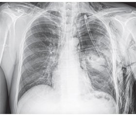
Актуальність. Травми грудної клітки при бойових діях посідають чільне місце й нерідко стають причиною смерті. До широкого впровадження методів візуалізації в клінічну практику рівень смертності при бойових травмах грудної клітки перевищував 50 %. Мета: порівняльна оцінка можливостей рентгенографії та ультрасонографії в діагностиці пневмотораксу, що виник у результаті бойової травми. Матеріали та методи. Проведено порівняльний аналіз результатів рентгенографії та ультрасонографії в 76 пацієнтів з пневмотораксом, що виник унаслідок бойової травми грудної клітки. Результати. При рентгенографії в положенні пацієнтів лежачи на спині чутливість методу становила 58,1 %, специфічність — 72,7 %, точність — 64,5 %, позитивна передбачувальна цінність — 73,5 %, негативна передбачувальна цінність — 57,1 %. Чутливість методу в положенні пацієнтів сидячи становила 71,9 %, специфічність — 89,5 %, точність — 76,3 %, позитивна передбачувальна цінність — 95,3 %, негативна передбачувальна цінність — 51,5 %. Ультразвукова діагностика пневмотораксу ґрунтувалася на виявленні симптому штрих-коду через відсутність ковзання вісцеральної плеври під час вдиху пацієнта. Чутливість ультрасонографії в В-режимі становила 90,8 %, специфічність — 81,8 %, точність — 89,5 %, позитивна передбачувальна цінність — 96,7 %, негативна передбачувальна цінність — 60,0 %, а в комбінованому В+М-режимі — 94,0; 88,9; 93,4; 98,4; 66,7 % відповідно. У діагностиці великого пневмотораксу чутливість рентгенографії становила 96,8 %, специфічність — 100,0 %, точність — 96,9 %, позитивна передбачувальна цінність — 100,0 %, негативна передбачувальна цінність — 50,0 %, а ультрасонографії — 96,7; 100,0; 96,9; 100,0; 66,7 % відповідно. Висновки. Ультрасонографія має більшу чутливість у діагностиці невеликого пневмотораксу, ніж звичайна рентгенографія, особливо в лежачих пацієнтів. Ультрасонографія в комбінованому В+М-режимі може бути як первинним, так і уточнюючим методом діагностики пневмотораксу при бойовій травмі.
Background. Chest injuries during combat operations occupy a prominent place and often become the cause of mortality. Before the widespread introduction of imaging methods into clinical practice, the mortality rate for chest combat injuries exceeded 50 %. Objective: a comparative assessment of radiography and ultrasonography options in the diagnosis of pneumothorax that occurred as a result of combat trauma. Materials and methods. A comparative analysis of the radiography and ultrasonography results was carried out in 76 patients with pneumothorax due to chest combat trauma. Results. During X-ray in the supine position, the sensitivity of the method was 58.1 %, specificity — 72.7 %, accuracy — 64.5 %, positive predictive value — 73.5 %, negative predictive value — 57.1 %. The sensitivity of the method in the sitting position of patients was 71.9 %, specificity — 89.5 %, accuracy — 76.3 %, positive predictive value — 95.3 %, negative predictive value — 51.5 %. Ultrasound diagnosis of pneumothorax was based on identifying the “barcode” sign due to the lack of sliding of the visceral pleura during the patient’s inspiration. The sensitivity of ultrasonography in B-mode was 90.8 %, specificity — 81.8 %, accuracy — 89.5 %, positive predictive value — 96.7 %, negative predictive value — 60.0 %, and in combined B + M modes — 94.0, 88.9, 93.4, 98.4, 66.7 %, respectively. In the diagnosis of large pneumothorax, the sensitivity of radiography was 96.8 %, specificity — 100.0 %, accuracy — 96.9 %, positive predictive value — 100.0 %, negative predictive value — 50.0 %, respectively, and of ultrasonography — 96.7, 100.0, 96.9, 100.0, 66.7 %, respectively. Conclusions. Ultrasonography has greater sensitivity for diagnosing small pneumothorax than conventional radiography, especially in bedridden patients. Ultrasonography in combined B + M modes can be both a primary and a clarifying method for diagnosing pneumothorax in combat trauma.
пневмоторакс; бойова травма; рентгенографія; ультрасонографія
pneumothorax; combat trauma; X-ray diagnosis; ultrasonography
Для ознакомления с полным содержанием статьи необходимо оформить подписку на журнал.
- 1. Singleton JA, Gibb IE, Bull AM, Mahoney PF, Clasper JC. Primary blast lung injury prevalence and fatal injuries from explosions: insights from postmortem computed tomographic analysis of 121 improvised explosive device fatalities. J Trauma Acute Care Surg. 2013;75(2 Suppl 2):S269-74.
- 2. Yakovenko VV, Grechanik EI, Abdullayev RYa, Bychenkov VV, Gumenyuk KV, Sobko IV. Modeling of the influence of fragments of ammunition on the biological tissue of a military in protective elements of combat equipment. Azerbaijan Medical Journal. 2020;4:107-115.
- 3. Durso AM, Caban K, Munera F. Penetrating thoracic Injury. Radiol Clin North Am. 2015;53(4):675-93.
- 4. Edgecombe L, Sigmon DF, Galuska MA, Angus LD. Thoracic trauma. In: StatPearls. Treasure Island (FL): StatPearls Publishing; May 29, 2022.
- 5. Schoenfeld AJ, Dunn JC, Bader JO, Belmont PJ Jr. The nature and extent of war injuries sustained by combat specialty personnel killed and wounded in Afghanistan and Iraq, 2003–2011. J Trauma Acute Care Surg. 2013;75(2):287-91.
- 6. Keneally R, Szpisjak D. Thoracic trauma in Iraq and Afghanistan. J Trauma Acute Care Surg. 2013;74(5):1292-7.
- 7. Lichtenberger JP, Kim AM, Fisher D et al. Imaging of combat-related thoracic trauma — review of penetrating trauma. Mil Med. 2018;183(3–4):e81-e88.
- 8. Klausner MJ, McKay JT, Bebarta VS et al. Warfighter personal protective equipment and combat wounds. Med J (Ft Sam Houst Tex). 2021(Pb 8-21-04/05/06):72-77.
- 9. Kong VY, Liu M, Sartorius B, Clarke DL. Open pneumothorax: the spectrum and outcome of management based on Advanced Trauma Life Support recommendations. Eur J Trauma Emerg Surg. 2015;41(4):401-404.
- 10. Tran J, Haussner W, Shah K. Traumatic Pneumothorax: a review of current diagnostic practices and evolving management. J Emerg Med. 2021;61(5):517-528.
- 11. Daurat A, Millet I, Roustan J-P, Maury C, Taourel P, Jaber S et al. Thoracic trauma severity score on admission allows to determine the risk of delayed ARDS in trauma patients with pulmonary contusion. Injury. 2016;47(1):147-53.
- 12. Tataroglu O, Erdogan ST, Erdogan MO et al. Diagnostic accuracy of initiaI chest x-rays in thorax trauma. J Coll Physicians Surg Pak. 2018;28(7):546-548.
- 13. Abdulrahman Y, Musthafa S, Hakim SY, Nabir S, Qan-bar A, Mahmood I et al. Utility of extended FAST in blunt chest trauma: is it the time to be used in the ATLS algorithm? World J Surg. 2015;39:172-8.
- 14. Langdorf MI, Medak AJ, Hendey GW et al. Prevalence and clinical import of thoracic injury identified by chest computed tomography but not chest radiography in blunt trauma: Multicenter prospective cohort study. Ann Emerg Med. 2015;66(6):589-600.
- 15. Moussavi N, Davoodabadi AH, Atoof F, Razi SE, Behnampour M, Talari HR. Routine chest computed tomogra-phy and patient outcome in blunt trauma. Arch Trauma Res. 2015;4(2):e25299.
- 16. Soult MC, Weireter LJ, Britt RC, Collins JN, Novosel TJ, Reed SF, Britt LD. Can routine trauma bay chest x-ray be bypassed with an extended focused assessment with sonography for trauma examination? Am Surg. 2015;81:336-40.
- 17. Ekpe EE. Overview of blunt chest injury with multiple rib fractures. Brit J of Medicine & Medical Research. 2016:12(8):1-15.
- 18. Pantea MA, Maev RG, Malyarenko EV, Baylor AE. A physical approach to the automated classification of clinical percussion sounds. J Acoust Soc Am. 2012;131(1):608-619.
- 19. Roberts DJ, Leigh-Smith S, Faris PD et al. Clinical presentation of patients with tension pneumothorax: a systematic review. Ann Surg. 2015;261(6):1068-1078.
- 20. Sabri YY, Hafez MAF, Kamel KM, Abbas DD. Evaluating the role of ultrasound in chest trauma: Common complications and computed tomography comparative evaluation. The Egyptian Journal of Radiology and Nuclear Medicine. 2018;49(4):986-992. https://doi.org/10.1016/j.ejrnm.2018.06.006.
- 21. Abdalla W, Elgendy M, Abdelaziz AA, Ammar MA. Lung ultrasound versus chest radiography for the diagnosis of pneumothorax in critically ill patients: A prospective, single-blind study. Saudi J Anaesth 2016;10:265-9. DOI: 10.4103/1658-354X.174906.
- 22. Attia YZ, Abd Elgeleel NM, El-Hariri HM, Ellabban GM, El-Setouhy M, Hirshon JM, Elbaih AH, El-Shinawi M. Comparative study of National Emergency X-Radiography Utilization Study (NEXUS) chest algorithm and extended focused assessment with sonography for trauma (E-FAST) in the early detection of blunt chest injuries in polytrauma patients. African Journal of Emergency Medicine. 2023;13:52-57. https://doi.org/10.1016/j.afjem.2023.02.003. PMID: 36937618. PMCID: PMC10014268. DOI: 10.1016/j.afjem.2023.02.003.
- 23. Avila J, Smith B, Mead T, Jurma D, Dawson M, Mallin M, Dugan A. Does the Addition of M-Mode to B-Mode Ultrasound Increase the Accuracy of Identification of Lung Sliding in Traumatic Pneumothoraces? Journal of Ultrasound in Medicine. 2018;37(11):2681-2687.

