Международный эндокринологический журнал 5 (69) 2015
Вернуться к номеру
Computer-Tomographic characterization of adrenal neoplasms
Авторы: H.А. Аlimukhamedova, Z.Yu. Khalimova - RSNPMC of Endocrinology, Таshkent, Uzbekistan
Рубрики: Эндокринология
Разделы: Клинические исследования
Версия для печати
Aim: to study computed tomographic (CT) characteristics of adrenal neoplasms.
Materials and methods
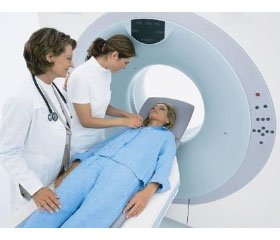
We examined 98 inpatients and outpatients with adrenal neoplasms, 51% and 49% of men and women, respectively (mean age 36.4 ± 1.29 years), followed up at the Center for Scientific and Clinical Study of Endocrinology, Uzbekistan Public Health Ministry. 22 patients with arterial hypertension without adrenal pathology matching by sex and age were included into a control group. The patients’ complaints were analyzed, complete clinical examination with measurement of body mass index (BMI) and arterial pressure was performed. Total and blood count and blood biochemical analysis were made with measurement of hormones. A 64-multislice computed tomography scanner in spiral mode (Brilliance 64 Phillips, Eindhoven, the Netherlands) was used to perform multi-slice spiral computed tomography.
Results and discussion
CT parameters of adrenal neoplasms were analyzed by their size. Thus, in patients with neoplasms with size of 1 cm (n=57) the adrenal gland length was 3.0 ± 0.26/3.06 ± 0.22 cm, that is, significantly more than in the controls (P<0.01 and P<0.001, on the right and on the left respectively). The adrenal gland thickness and height were 1.5-2 times more than those in the controls not differing from those in patients with neoplasms with size more than 1 cm. Densitometric parameters of the neoplasms ranged from 27.1 ± 1.59 to 34.86 ± 2.2 on the left and from 25.3 ± 1.32 to 38.13 ± 3.06 on the right, mean 39.1 ± 1.87 HU. The neoplasms were dense; their density was lower than the one of those with size of more than 3 cm, exceeding similar parameters in the controls by 2.4 times. Moreover, they were non-homogenous and had various densities at various regions.
The length, thickness and height of the adrenals in patients with neoplasms of size ranging from 1 to 3 cm (n=17) significantly differed from those in the controls not differing from those in other two groups. Despite large sizes of the neoplasms, their density was lower than those in patients in the 1st group, sometimes reaching normal densitometric values mimicking disintegration and necrosis of a tumor. Their density parameters ranged from 27.4 ± 3.14 to 40.8 ± 3.68 HU, mean value was 34.1 ± 3.62 HU.
Density of neoplasms in patients with large adrenal neoplasms (n=24) were significantly higher than the one in the controls and patients of other two groups. The neoplasms originated from pathologically changed hyperplastic adrenal tissue with typical thickening of the adrenal pedicles and increasing of length to 3.1 ± 0.38 cm.
To study association between CT-picture of the adrenal neoplasms and hormonal status the parameters above were examined separately by hormonal deviations. There were the following nosologic forms of adrenal tumors in the patients examined: pheochromocytomas (9.2%, n=9), aldosteromas (11.2%, n=11), corticosteromas (21.4%,n=21) and hormonally inactive tumors (58.2%, n=57).
In patients with corticosteromas CT-picture was a peculiar one. It should be emphasized that in persons with normal cortisol the neoplasm’s density is significantly different as compared with those with hypercortisolemia (43.4 ± 0.7 HU versus 36.5 ± 4.1 HU, P<0,05); there were differences in the adrenal tissue’s density variation and increase in the adrenal gland length, thickness and height. Thus, contrary to patients with normal cortisol corticosteromas were characterized with significant relative reduction in the density of neoplasms, density and size of the adrenals
Next, parameters of patients with aldosteromas and those with normal aldosterone were compared. The findings demonstrated that contrary to parameters in the patients with adrenal neoplasms but with normal secretion of aldosterone those in patients with aldosteromas are different from those in the controls and those in patients with normoaldosteronemia. In persons with aldosteromas the adrenals were larger in size, their density variation prevailed, and density of the neoplasm was a peculiar one.
Excluding significant deviations in length, thickness and height of the adrenals, neoplasms in patients with pheochromocytomas were poorly distinguishable by density. These neoplasms buried in moderately hyperplastic adrenal tissue having significantly lower density than those in patients with the adrenal neoplasms and normal catecholamines in the daily urine.
Conclusions
Computed tomography allows assessing changes in length, thickness and height of the adrenal glands (Р<0.01 and Р<0.001 in the left and in the right, respectively), densitometric parameters (P<0.001) as well as the gland’s structure, neoplasm’s size, and hormonal activity which are significantly different from those in the healthy controls. CT picture of the adrenal neoplasms depends not only on a neoplasm’s size but also on functional state of its cell structure. The adrenals neoplasms are characterized with the increase in density depending on the neoplasm’s size and always originating in the primarily changed adrenal tissue with diffuse or focal hyperplasia; the increase in the adrenal’s size, differences in densitometric parameters as compared with normal ones from normal to maximum.

